Figures & data
Figure 1. PMTA of uterine myoma was performed in a patient and the microwave antenna (A) and thermocouple needle (T) were inserted percutaneously into the target.
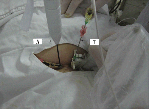
Figure 2. (A) The ultrasound image of 2D grey-scale of uterus myoma at 10 s after MW therapy. Echo enhancement began around the antennas. (B) Ultrasound image of 2D grey-scale of uterus myoma at 70 s after MW therapy. The area of echogenicity is noted to be enlarged. (C) Ultrasound image of 2D grey-scale of uterus myoma at the end of ablation. The whole myoma is obscured due to echoes.
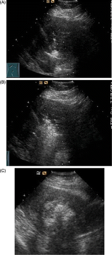
Figure 3. (A) CDFI of myoma before ablation shows the myoma with hyperperfusion. (B) CDFI of ablated myoma at the end of ablation. No residual colour Doppler is demonstrated.
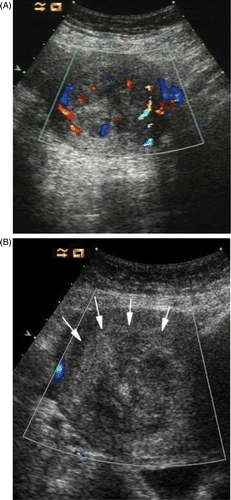
Figure 4. (A) Contrast enhanced ultrasound (ceUS) image of myoma before ablation. The fibroid was obviously enhanced. (B) CeUS of myoma 1 day following ablation showing no ultrasound contrast enhancement in the myoma.
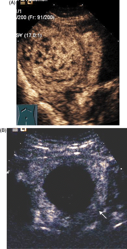
Figure 5. (A) CeMRI picture of myoma before ablation shows the fibroid was obviously enhanced. (B) Sagittal ceMRI of the pelvis following PMTA of a fibroid in the posterior wall of uterus. No enhancement is seen in the fibroid.
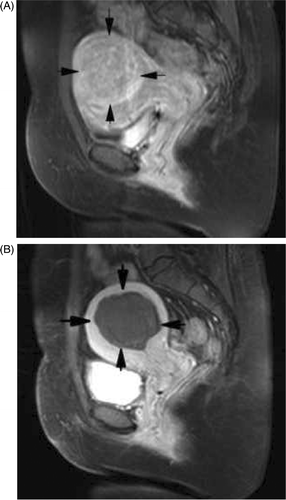
Table I. Fibroid volume and volume reduction at baseline, 3, 6, 9 and 12 months follow up.
Table II. Patient symptoms after ablation.