Figures & data
Figure 1. Transverse contrast-enhanced CT scans in a 55-year-old woman with ovarian carcinoma metastasis. (A) Preablation scan showed one hypoattentuating neoplasm (arrows) near the hilum. (B) Scan obtained 9 months after the first ablation shows hypoattenuating ablation zone (arrows) without enhancement. The needle track was cauterized as the probe was removed, resulting in the subcapsular triangle-shaped thermal lesion (arrowheads). (C) Scan obtained 32 months after the second ablation shows hypoattenuating ablation zone (arrows) without enhancement corresponding to treated region.
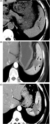
Figure 2. Transverse contrast-enhanced CT scans in the 56-year-old woman with lung adenocarcinoma metastasis. (A) Preablation scan showed one solitary neoplasm (arrow) with low attenuation and peripheral enhancement at the lower pole of the spleen. (B) On an arterial phase CT scan obtained 28 months after treatment no enhancement is seen in the enlarged coagulation zone (arrows), suggesting the absence of residual tumour.

Table I. Clinical features and outcomes of the four patients.