Figures & data
Figure 1. Geometry of the treatment domain. The origin is located in the centre of the transducer's face. The geometric focus of the transducer is at 13 cm and the water filled distance from the transducer to the skin is 11.4 cm (11.0 for some simulations, as measured from the centre of the face of the transducer). A small (1.1 cm3) and medium (2.6 cm3) volume tumour were studied with this geometry. A deeper medium and a larger (4.5 cm3) tumour were studied with a slightly modified geometry using an expanded simulation region.
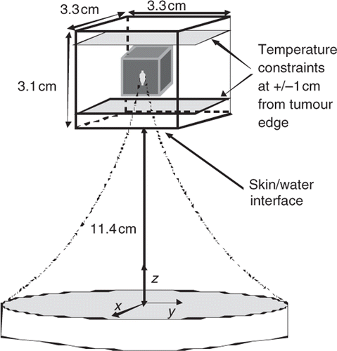
Figure 2. Treatment time vs. perfusion for three focal zone paths treating the small superficial tumour with 45 positions with the standard total applied power and a range of perfusions. Also shown are the percentages of the treatment time spent cooling the normal tissue. The lines are for the AS (MBF), XY (Ra) (squares); 3D Kn (circles) and PL (BMF), XY Ra (triangles) scan patterns.
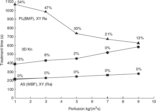
Table I. Thermal and acoustic properties used in all simulations Citation[1], Citation[2].
Table II. Simulation configurations. All distances are given in cm from the transducer's centre, the transducer's geometric focus is at 13 cm, and all dimensions are in cm. The coordinate order is X, Y, Z.
Figure 3. The successive focal zone locations for: (Left) the ‘planar (BMF); XY raster’ path, in which the back plane is treated first with an X-Y raster scan, then the middle plane and then the front plane; (Right) the ‘axially stacked (MBF), XY raster scan’ in which the transducer is fixed at one transverse (X-Y) location and the focal zone is electronically scanned through the stack of three axial M,B and F locations. The transducer is then moved through the remaining transverse locations in an XY raster pattern, applying the same axial stack order at each location. The numbers specify the order in which the focal zone positions were scanned.
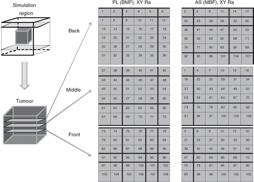
Figure 4. Axial view of four transverse, XY paths used to scan an axial stack: inner middle outer (IMO), concentric annuli (top left), knight (top right), adjacent square annuli (bottom left), and adjacent rectangular annuli (bottom right).
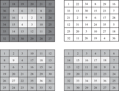
Table III. Scanning patterns studied: F indicates the front position in an axial stack, or the front plane in a planar scan; M and B indicate the middle and back. MFB indicates that the M position (or plane) was treated first, etc.
Figure 5. Treatment time vs. scanning path type for the medium superficial tumour (108 focal zone positions) and the standard treatment conditions. Also shown are the percentages of the treatment time spent cooling the normal tissue. When the cooling time percentages are nearly identical, only one value (located between the paths with similar values) is shown.

Figure 6. Treatment time vs. power density for three scan paths treating the small superficial tumour with 45 positions. The standard perfusion and constraint values were used but the total applied power was varied. Also shown are the percentages of the treatment time spent cooling the normal tissue. The lines are for the AS (MBF), XY (Ra) (squares); 3D Kn (circles) and PL (BMF), XY Ra (triangles) scan patterns.
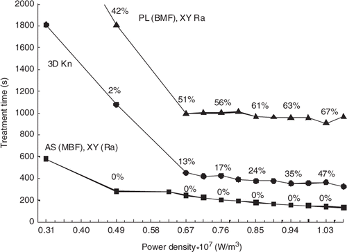
Figure 7. Pulse heating and inter-pulse cooling times (s) during treatment of the medium, superficial tumour under the standard treatment conditions: (A) The heating times at each sequential focal zone position for both the axially stacked (MBF), XY raster path (squares) and the planar (BMF), XY raster path (x). (B) The cooling times at each sequential focal zone position for the planar (BMF), XY raster path. The cooling times for the axially stacked (MBF), XY raster are not shown because they are zero for almost all positions in the treatment and are only a few seconds for the remaining positions.
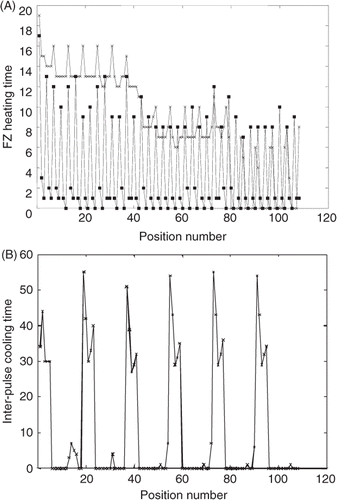
Figure 8. Temperature vs. time for the middle, front, back, and transversely adjacent (to either side of the middle position in the plane perpendicular to the transducer axis) locations with respect to the FZ location when the beam is focused on the middle (M) position. Results are shown for the first heating pulse for the larger tumour under standard treatment conditions.
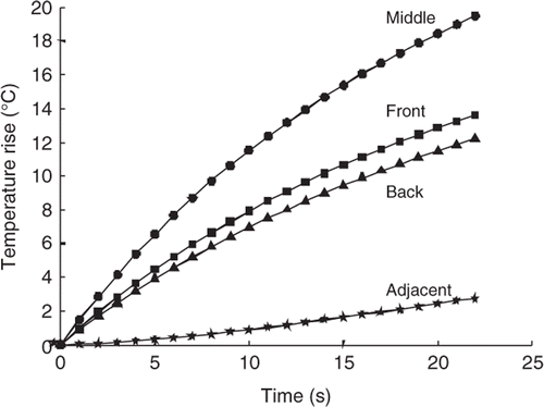
Figure 9. Treatment time vs. transverse scan path for axially stacked (MBF) stacks treating the medium, superficial tumour for 6°C and 5°C normal tissue temperature limits under the standard treatment conditions. The scans are rank ordered based on their times for the 5°C constraint case. The path abbreviations are: Kn for the AS (MBF) XY knight jumps; inner I, middle M, outer O for the AS (MBF) concentric annuli; Ra for the AS (MBF) XY raster; Rec for the AS (MBF) adjacent rectangular annuli; and Sq for the adjacent square annuli. Paths where the treatment times were extrapolated have an asterisk. The percentages of treatment time spent cooling the normal tissue for each path with the 5°C limit are shown. The percentages for the 6°C limit are not shown because they were all less than 3% and were not a significant factor in determining total treatment time.
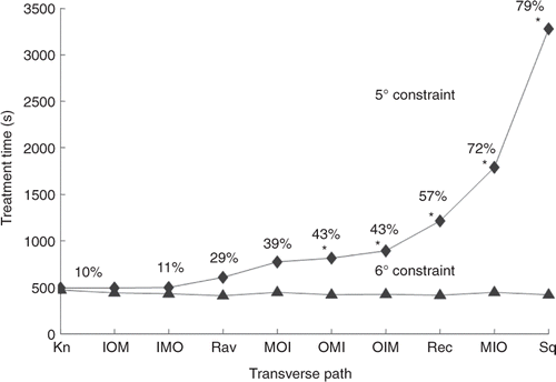
Figure 10. Treatment time (s) vs. transverse path for (A) the medium deep (top) and (B) larger tumour (bottom) for the standard treatment conditions and a NT temperature limit of 8°C.The independent single pulse treatment times for these tumours are 3852 and 2556 s, respectively (Appendix). The percentage of treatment time spent cooling is shown above each path (or between points in cases where the percentages were essentially identical).
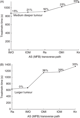
Figure 11. The stack heating time vs. stack number for the paths in . The graphs, in order from shortest to longest treatment time, are for the AS (MFB); (A) raster, (B) IMO concentric annuli, (C) IOM concentric annuli, (D) OMI concentric annuli, and (E) knight. Cooling times of greater than 30 s in a preceding stack are marked with a tilde.
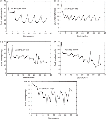
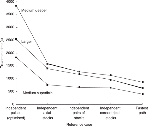
Figure 12. Treatment time vs. reference case for the medium superficial, medium deeper, and larger tumour treated with ‘independent’ pulses and stacks under the standard treatment conditions. Also shown are the times for the fastest scans obtained in this study for the same treatment conditions. The fastest case for each of the tumours was; AS (MFB) XY raster for both the medium superficial tumour and for the larger tumour, and AS, MFB IMO concentric annuli for the medium deeper tumour.