871
Views
46
CrossRef citations to date
0
Altmetric
Research Article

Ultrasound-guided high intensity focused ultrasound treatment for extra-abdominal desmoid tumours: Preliminary results

, , MD &
Pages 648-653 | Received 03 Apr 2011, Accepted 11 Jun 2011, Published online: 28 Jul 2011

Figures & data

Table I.  Patient and tumour characteristics.

Figure 1. A 7-year-old girl with multiple recurrent desmoid tumour nodules in the left leg and buttock, who received three surgical resections before HIFU ablation. Further resection was considered impossible. HIFU ablation was performed with a palliative aim. (A) Before HIFU ablation, there were multiple tumour nodules (arrows) which showed high enhancement on coronal T1-weighted contrast-enhanced MRI. (B) One month after HIFU ablation, almost all treated tumours (arrows) showed no enhancement. (C) Nine months after HIFU ablation, the treated area (arrow) showed no enhancement and shrank significantly. (D) Sixteen months after HIFU ablation, the treated area (arrow) showed no enhancement and shrank further.
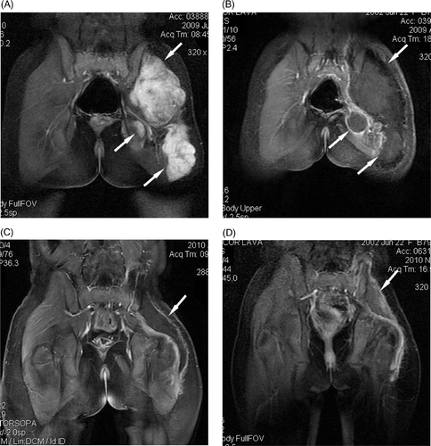
Figure 1. A 7-year-old girl with multiple recurrent desmoid tumour nodules in the left leg and buttock, who received three surgical resections before HIFU ablation. Further resection was considered impossible. HIFU ablation was performed with a palliative aim. (A) Before HIFU ablation, there were multiple tumour nodules (arrows) which showed high enhancement on coronal T1-weighted contrast-enhanced MRI. (B) One month after HIFU ablation, almost all treated tumours (arrows) showed no enhancement. (C) Nine months after HIFU ablation, the treated area (arrow) showed no enhancement and shrank significantly. (D) Sixteen months after HIFU ablation, the treated area (arrow) showed no enhancement and shrank further.

Figure 2. A 28-year-old woman with an abdominal wall desmoid tumour receiving no treatment before HIFU ablation. (A) Before HIFU ablation, the tumour (arrow) showed high enhancement on transverse T1-weighted contrast-enhanced MRI. (B) Four days after HIFU ablation, the treated area (arrow) showed no contrast enhancement, the skin overlying the treated area was swollen. (C) Ten months after HIFU ablation, no contrast enhancement was observed in the treated area (arrow), which shrank significantly. (D) Eighteen months after HIFU ablation, no enhancement was observed in the treated area (arrow), which shrank further.
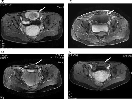
Figure 2. A 28-year-old woman with an abdominal wall desmoid tumour receiving no treatment before HIFU ablation. (A) Before HIFU ablation, the tumour (arrow) showed high enhancement on transverse T1-weighted contrast-enhanced MRI. (B) Four days after HIFU ablation, the treated area (arrow) showed no contrast enhancement, the skin overlying the treated area was swollen. (C) Ten months after HIFU ablation, no contrast enhancement was observed in the treated area (arrow), which shrank significantly. (D) Eighteen months after HIFU ablation, no enhancement was observed in the treated area (arrow), which shrank further.

Reprints and Corporate Permissions

Please note: Selecting permissions does not provide access to the full text of the article, please see our help page How do I view content?

To request a reprint or corporate permissions for this article, please click on the relevant link below:

Academic Permissions

Please note: Selecting permissions does not provide access to the full text of the article, please see our help page How do I view content?

Obtain permissions instantly via Rightslink by clicking on the button below:

If you are unable to obtain permissions via Rightslink, please complete and submit this Permissions form. For more information, please visit our Permissions help page.