Figures & data
Figure 1. Schematic diagram of the progress of the coagulation zone in the tissue during radiofrequency ablation. At the beginning, the coagulated tissue is mainly located at the edges (distal and proximal), while at the end of heating the coagulated lesion has broadly extended and forms an ellipsoidal shape with the longest axis being the electrode axis. The bold lines represent the tissue heated over 100°C, i.e. strongly dehydrated zones.
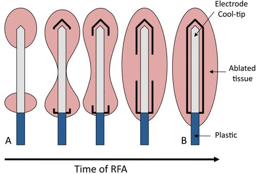
Figure 2. Placement of the multi-thermocouple probe tied to the electrode. T1, T2 and T3 are the locations of the thermocouples (dimensions in mm).
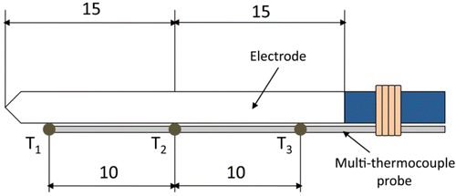
Figure 3. Experimental set-up to record temperatures and impedance evolution. The multi-thermocouple probe and electrode were jointly inserted into the tissue. The electrode was internally cooled by a peristaltic pump.
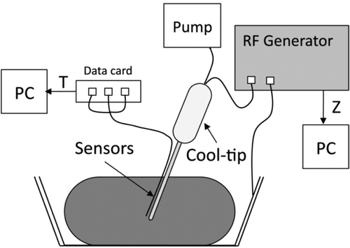
Figure 4. Geometry of the two-dimensional theoretical model (out of scale, and dimensions in mm). The domain is divided into three zones: plastic portion of the electrode, metallic electrode, and hepatic tissue.
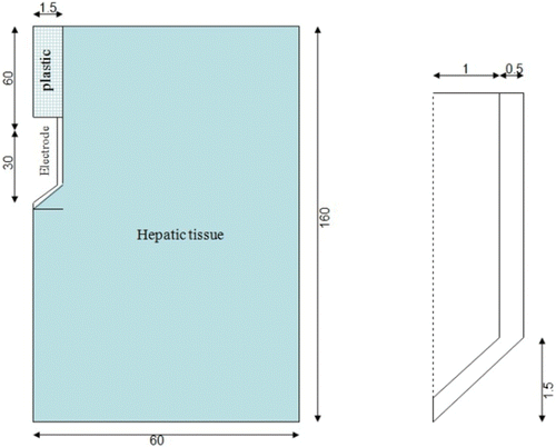
Figure 5. Change in the tissue thermal and electrical conductivity with temperature.
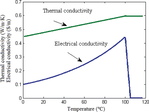
Table I. Characteristics of the materials used in the model Citation14, Citation15.
Figure 6. Results from ex vivo experiments and theoretical model. Mean temperature evolution of the sensors placed in the tissue surrounding the surface of the electrode tip: T1, T2 and T3 were the thermocouples placed at 0.5, 1.5 and 2.5 mm, respectively, from the electrode tip. Mean progress of the impedance (Z exp). Impedance progress computed from the theoretical model (Z theor). Note that the impedance began increasing when temperature at the middle of the electrode (T2) became approximately steady at around 50 s.
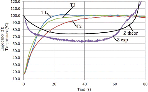
Figure 7. Results from ex vivo experiments. Progress of the impedance (Z) and temperature evolution of the sensors placed in the tissue surrounding the surface of the electrode tip: T1, T2 and T3 were the thermocouples placed at 0.5, 1.5 and 2.5 mm, respectively, from the electrode tip. Dots are mean values and error bars are standard deviations.
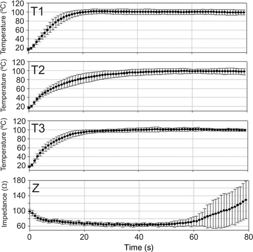
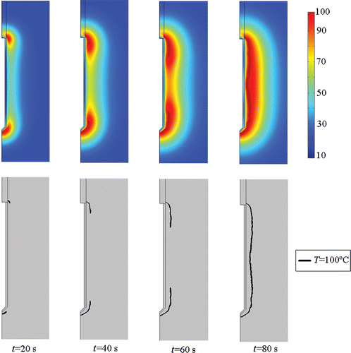
Figure 8. Temperature distributions computed from the theoretical model (top, scale in °C) and progress of the 100°C isotherm in the tissue (bottom). Note that the 100°C isotherm completely encloses the electrode at ≈80 s.