Figures & data
Figure 1. Clinical and histologic appearance of neck lesions in a 35-year-old man with warts and Darier disease. (a) Warts (cycled) superimposed on lesions of Darier disease. (b) Diffuse vacuolated keratinocytes in the upper epidermis were characteristic of HPV-infected skin; epidermal invagination and suprabasal lacunae with dyskeratotic acantholytic cells were characteristic of Darier disease (hematoxylin-eosin, frozen section of neck lesion, original magnification ×200). (c) DNA segment specific for HPV type 6. (d) At 15 days after hyperthermia treatment, all papules and plaques in the neck disappeared completely, but Darier disease persisted.
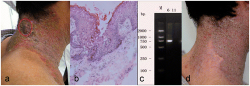
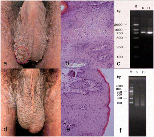
Figure 2. Clinical and histologic appearance of scrotal lesions in a 35-year-old man with warts and Darier disease. (a) Papillary and cauliflower-shaped papules and plaques in the scrotum. (b) Patches of vacuolated keratinocytes in thickened epidermis were characteristic of HPV-infected skin (hematoxylin-eosin, original magnification ×200). (c) DNA segments specific for HPV types 6 and 11. (d, e) Clinical and histologic findings after 2 months hyperthermia treatment, showing resolution of the warty lesions. (f) No HPV-specific DNA was detected after 2 months treatment.