Figures & data
Table 1. Comparison of RFA and cryoablation in terms of total ablated volume.
Figure 1. Schematic diagram of a renal tumour and tumour margin according to the location of the tumour. A parenchymal tumour (A) has a greater volume of tumour margin than an exophytic (B) or central (C) tumour in cases where all the tumours are the same size. The parenchymal tumour requires double the ablation of the tumour margin than an exophytic or central tumour that is projecting 50% from the renal parenchyma to the peri-renal space or renal sinus. RP indicates renal parenchyma.
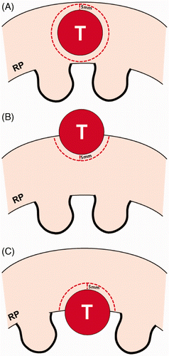
Figure 2. Schematic diagram of a renal tumour and tumour margin according to the number of tumours. (A) An exophytic tumour is projecting 50% out from the renal parenchyma. RP indicates renal parenchyma. (B) There are three tumours consisting of one parenchymal tumour (left) and two exophytic tumours, all the same size as figure A. These exophytic tumours are projecting 70% (middle) and 30% (right) from the parenchyma into the peri-renal space, respectively. Renal tumours in figure B require four times the normal renal tissue for thermal ablation than in figure A.
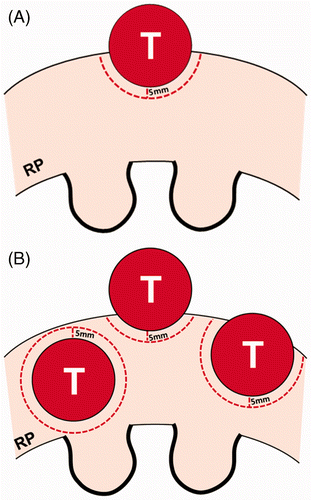
Figure 3. Over-ablation beyond the tumour margin during the RFA procedure. Arrowheads indicate RFA electrodes. (A) For radiofrequency ablation (RFA) of a spherical RCC (red), the shape of ablation zone (yellow) is not a circle but oval. Thus, the lateral areas of the RCC and tumour margin (dotted line) require additional ablations to prevent a residual or recurrent lesion. (B and C) Additional ablations (blue and violet) are performed to completely cover the tumour margin (dotted line) as well as the RCC (red). However, some over-ablation of normal tissue is created beyond the lateral tumour margin.
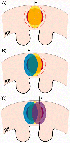
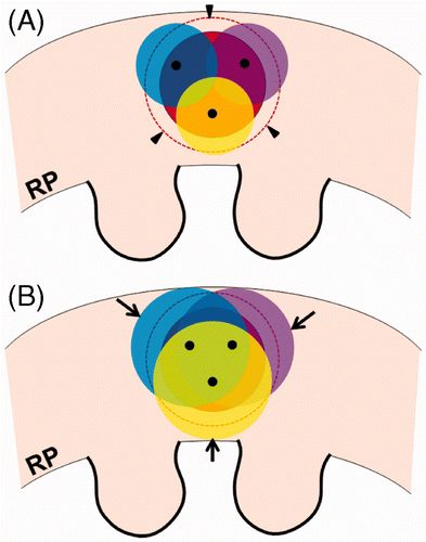
Figure 4. Over-ablation beyond the tumour margin during the cryoablation procedure. (A) For cryoablation of a spherical RCC (red), the ends (black dots) of three cryoprobes are seen within the tumour. The ice balls (yellow, blue, and violet) do not cover the entire tumour margin (arrowheads). (B) The ice balls grow to completely cover the tumour margin. However, some over-ablation of normal renal tissue (arrows) is noted beyond the tumour margin.