Figures & data
Figure 1. Physiological response to 5-day exposure to moderate hyperthermia. Mice were implanted with intraperitoneal thermal sensors, recovered for 7 days, and core temperature (A, B), heart rate (C, D), and activity level (E, F) in male (A, C, E) and female (B, D, F), respectively, were continuously monitored during 5-day exposure to either 22°C (NT) or 37°C (cHT) ambient temperature. Core temperature and heart rate were measured every 20 s and 1-h means calculated. Activity was measured every 20 s and 4-h means calculated. Four experiments, each with four mice of each sex per group, were pooled. Data are presented as means ± SEM. The differences between NT and cHT mice for each sex are indicated on the figures. NT female mice maintained a 0.55°C higher core temperature than NT males (p = 0.005), but cHT female mice maintained 1.2°C lower core temperature than cHT males (p = 0.002). There were no significant differences in heart rate or activity level between the two sexes.
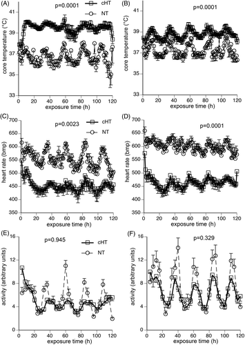
Table I. Change in weight, water and food intake, and survival during 5-day exposure to Normothermic or Hyperthermic conditions1.
Figure 2. Effect of 5-day chronic moderate heat exposure on expression of heat shock proteins in lung and brain. Male and female mice exposed to moderate hyperthermia for 5-days (cHT) and normothermic (NT) control mice were euthanised, the lungs (A, B) and brain (C, D) snap-frozen, and tissue homogenates analysed for Hsp25, Hsp72, and Hsp90 levels by immunoblotting. HSP band densities were expressed as ratio to ß-tubulin band densities and standardised to NT male levels for each experiment. (A, C) Mean ± SEM of 16 female and 13 male mice are displayed. (B, D) Representative blots. *, †, and ¶ indicate p < 0.05 versus NT male, cHT female, and NT female, respectively.
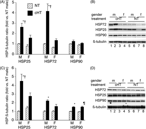
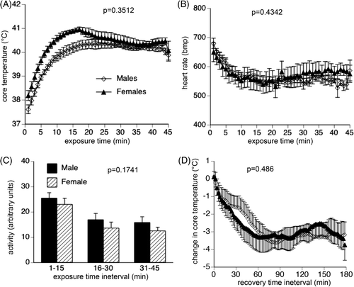
Figure 3. Physiological response to acute exposure to uncompensable heat stress. Following implantation of intraperitoneal thermal sensors, male and female mice with were exposed to 43°C for 45 min, then returned to 22°C while continuously monitoring core temperature, heart rate and activity level from the intraperitoneal sensors. Core temperature (A) and heart rate (B) were measured every 20 s, and 1-min means calculated. Activity (C) was measured every 20 s, and 15-min means calculated. (D) During recovery, core temperature was measured every 20 s, 2-min means calculated and expressed as change from the core temperature measured at the end of the acute heat exposure. Data are expressed as mean ± SEM of 16 male and 16 female mice. P values are indicated in the figure panels.