Figures & data
Figure 1. Caspase-3 level after DOX treatment. n = 3 experiments. Data were presented as mean ± SD. *Statistical significance comparing each treatment to control cells (cells without treatment), with one-sided Student's t-test (α = 0.05). ★Statistical significance among the groups by ANOVA with Bonferroni post-hoc correction (α = 0.05).
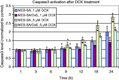
Figure 2. Hoechst/PI staining of MES-SA cells (upper panel) and Dx5 cells (lower panel) treated by incubator hyperthermia. Images were taken either immediately, or 24 h after the treatment. The scale bar represents 8 µm. (A) Control MES-SA; (B) MES-SA immediately after 1 h 43°C incubation; (C) MES-SA 24 h after 1 h 43°C incubation; (D) MES-SA immediately after 30 min 50°C incubation; (E) MES-SA 24 h after 30 min 50°C incubation; (F) control Dx5; (G) Dx5 immediately after 1 h 43°C incubation; (H) Dx5 24 h after 1 h 43°C incubation; (I) Dx5 immediately after 30 min 50°C incubation; (J) Dx5 24 h after 30 min 50°C incubation.
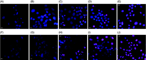
Figure 3. Hoechst/PI staining of MES-SA cells (upper panel) and Dx5 cells (lower panel) treated by laser excited (3 min) ICG (10 µM). Images were taken either 1 h after, or 24 h after the treatment. The scale bar represents 8 µm. (A) Control MES-SA; (B) MES-SA 1 h after laser irradiation; (C) MES-SA 24 h after laser irradiation; (D) MES-SA 1 h after laser irradiation; (E) MES-SA 24 h after laser irradiation ; (F) control Dx5; (G) Dx5 1 h after laser irradiation; (H) Dx5 24 h after laser irradiation; (I) Dx5 1 h after laser irradiation; (J) Dx5 24 h after laser irradiation.
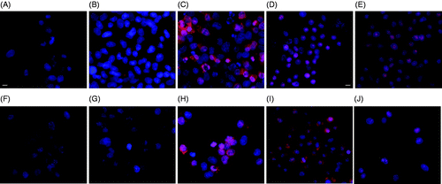
Figure 4. DOX fluorescence in MES-SA cells treated by only DOX (A and F), DOX + 1 h 43°C incubation (B and G), DOX + 30 min 50°C incubation (C and H), DOX + 3 min laser/5 µM ICG (D and I), and DOX + 3 min laser/10 µM ICG incubation (E and J). Images were taken either 1 h after (A, B, C, D and E), or 24 h after the treatment (F, G, H, I and J). The scale bar represents 8 µm.
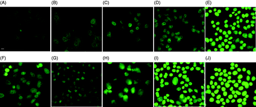
Figure 5. DOX fluorescence in Dx5 cells treated by only DOX (A and F), DOX + 1 h 43°C incubation (B and G), DOX + 30 min 50°C incubation (C and B), DOX + 3 min laser/5 µM ICG (D and I), and DOX + 3 min laser/10 µM ICG incubation (E and J). Images were taken either 1 h after (A, B, C, D and E), or 24 h after the treatment (F, G, H, I and J). The scale bar represents 8 µm.
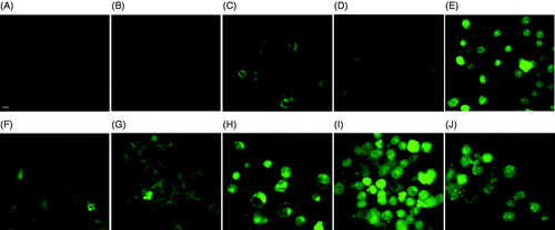
Figure 6. Hsp70 expression after different types of hyperthermia treatment (n = 3). Data were normalised as described above. ★Significantly higher cellular Hsp70 level among the three groups by ANOVA test with Bonferroni post-hoc correction.
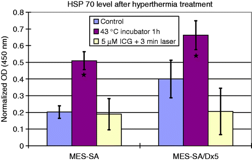
Figure 7. Calcein fluorescence after hyperthermia treatment. (A) MES-SA without any treatment; (B) MES-SA treated by 1 h 43°C incubator hyperthermia; (C) MES-SA treated with verapamil. Exposure time 1000 ms. The scale bar in (A) represents 24 µm.
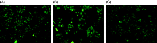
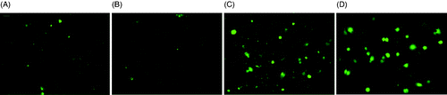
Figure 8. Calcein fluorescence after hyperthermia treatment. (A) Dx5 cells without any treatment; (B) Dx5 cells treated by 1 h 43°C incubator hyperthermia; (C) Dx5 cells treated by 5 µM ICG + 3 min laser; (D) Dx5 treated with verapamil. All the images were taken under 20× magnification and the exposure time was 1000 ms. The scale bar represents 24 µm.