2,173
Views
48
CrossRef citations to date
0
Altmetric
Review Articles

Protein interactomes of three stress inducible small heat shock proteins: HspB1, HspB5 and HspB8

&
Pages 409-422 | Received 26 Feb 2013, Accepted 02 Apr 2013, Published online: 22 May 2013

Figures & data

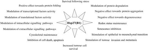
Figure 1. Cellular functions of HspB1. In addition to its well-known ability to protect cells against heat shock and other types of injuries, constitutively expressed HspB1 plays a major role in many different cellular processes, such as those listed in the figure.

Figure 1. Cellular functions of HspB1. In addition to its well-known ability to protect cells against heat shock and other types of injuries, constitutively expressed HspB1 plays a major role in many different cellular processes, such as those listed in the figure.

Table I. HspB1 interactome.

Figure 2. Native size and phosphorylation of HspB1 and structure-specific interaction with client protein targets. HeLa cells were lysed and the 10 000 × g cytosolic fraction containing all the cellular content of HspB1 was analysed by gel filtration column as previously described [Citation27]. Immunoblot analysis of two-by-two pooled fractions was performed using antibodies that are specific to either total HspB1 or phosphorylated (phospho-Ser15, phospho-Ser78 or phospho-Ser82) HspB1. The presence of three client proteins that interact with HspB1 was detected using specific antibodies recognising Pro-caspase-3, HDAC6 and STAT2. Three native size fractions could be defined depending on HspB1 phosphorylation: 50–200 kDa, phosphorylation at the level of serines 15 and 82, 200–400 kDa, phosphorylation at the level of serine 78 and 400–700 kDa oligomers containing phosphorylated serine 82. Note that pro-caspase-3 co-eluted mainly with the serine 15 phosphorylated small oligomers. HDAC6 was at the level of the large serine 82 phosphorylated oligomers while STAT2 had a less defined elution profile between the medium and large sized oligomers. Interactions of these proteins with different phospho-oligomeric structures of HspB1 was confirmed by co-immunoprecipitation [Citation44].

Figure 2. Native size and phosphorylation of HspB1 and structure-specific interaction with client protein targets. HeLa cells were lysed and the 10 000 × g cytosolic fraction containing all the cellular content of HspB1 was analysed by gel filtration column as previously described [Citation27]. Immunoblot analysis of two-by-two pooled fractions was performed using antibodies that are specific to either total HspB1 or phosphorylated (phospho-Ser15, phospho-Ser78 or phospho-Ser82) HspB1. The presence of three client proteins that interact with HspB1 was detected using specific antibodies recognising Pro-caspase-3, HDAC6 and STAT2. Three native size fractions could be defined depending on HspB1 phosphorylation: 50–200 kDa, phosphorylation at the level of serines 15 and 82, 200–400 kDa, phosphorylation at the level of serine 78 and 400–700 kDa oligomers containing phosphorylated serine 82. Note that pro-caspase-3 co-eluted mainly with the serine 15 phosphorylated small oligomers. HDAC6 was at the level of the large serine 82 phosphorylated oligomers while STAT2 had a less defined elution profile between the medium and large sized oligomers. Interactions of these proteins with different phospho-oligomeric structures of HspB1 was confirmed by co-immunoprecipitation [Citation44].

Table II. HspB5 interactome.

Table III. HspB8 interactome.
Reprints and Corporate Permissions

Please note: Selecting permissions does not provide access to the full text of the article, please see our help page How do I view content?

To request a reprint or corporate permissions for this article, please click on the relevant link below:

Academic Permissions

Please note: Selecting permissions does not provide access to the full text of the article, please see our help page How do I view content?

Obtain permissions instantly via Rightslink by clicking on the button below:

If you are unable to obtain permissions via Rightslink, please complete and submit this Permissions form. For more information, please visit our Permissions help page.