Figures & data
Figure 1. (A) MR-compatible positioning system; the transducer is at the centre of the image and a manual rotation and head--foot translation of the transducer is allowed by a dedicated chariot and a junction. Tooth and ear bars provide rigid stereotaxy under the transducer. (B) Full MRgFUS therapy set-up.
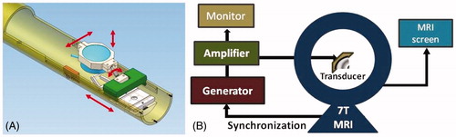
Table I. Characteristics of the different transducers studied.
Figure 2. T2 weighted images of the transducers on top of the rat head with the position and predicted extent of the corresponding focal spots. (A) Single element F/D = 1. (B) Single element F/D = 0.8.
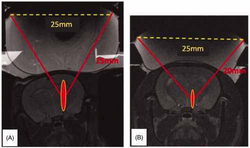
Figure 3. Brain tumour model in rat (RG2 glioma) and its kinetic: contrast enhanced T1-weighted MR images. Enlarged, day 12, is highlighted the growth time/volume chosen for treatments.
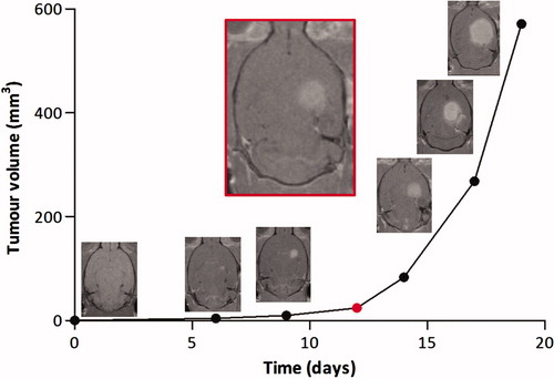
Table II. In vivo experiments at 1.5 MHz (temperature rise at the focus estimated from MR thermometry data).
Figure 4. Effect of water cooling: MR thermometry in the focal and cortex planes.
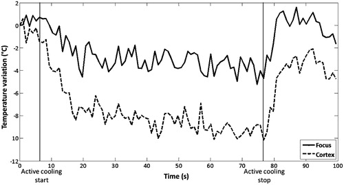
Table III. In vivo experiments at 1.5 MHz F/D = 0.8 (temperature rise at the focus estimated from MR thermometry data).
Figure 5. Different heating strategies with the 2.5 MHz transducer. (A) Single sonication at 4.33 MPa for 20 s. (B) Four sonications at 7.58 MPa, 2 s on 2 s off. (C) Two sonications at 7.58 MPa, 5 s on 5 s off. (D) Single sonication at 7.58 MPa for 7 s.
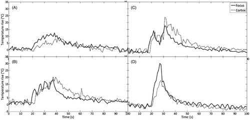
Table IV. In vivo experiments at 2.5 MHz F/D = 0.8 (temperature rise at the focus is estimated from MR thermometry data); 1–6 rats without tumours, 7–12 rats with tumours.
Table V. Summary of in vivo experiments.
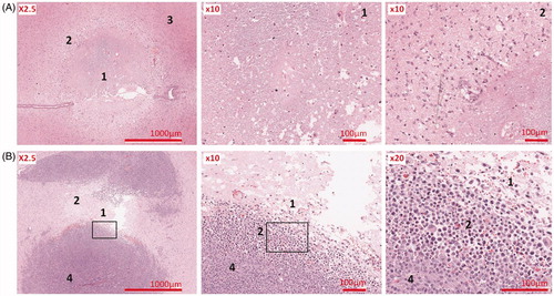
Figure 6. Image correlations. (A) Thermal lesion in the healthy brain. (B) Thermal lesion in the brain tumour. The red arrows indicate the region of acoustic energy deposition. The black arrows indicate the tumour location. (1) T2-weighted MRI, (2) MR-ARFI before treatment, (3) MR thermometry map during treatment, (4) T2-weighted MRI after treatment, (5) macroscopic section of whole brain H&E.
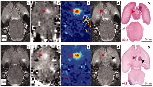
Figure 7. (A) Thermal lesion in healthy brain (rat sacrificed 48 h after HIFU); (B) Thermal lesion in brain tumour (rat sacrificed 4 h after HIFU). (1) centre, (2) peripheral, (3) healthy brain, (4) untreated tumour.