Figures & data
Figure 1. Physical set up and components of 3D visualisation preoperative treatment planning system. (1) A control unit of the electromagnetic tracking system, (2) two sensor interface devices of the electromagnetic tracking system, (3) a field generator of the electromagnetic tracking system, (4) tracked ultrasound probe, (5) a simulation model, (6) screen.
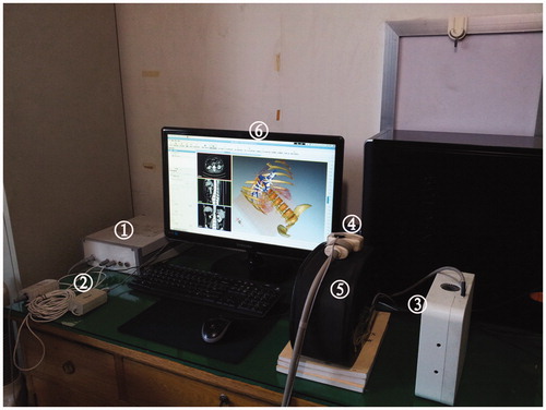
Figure 2. The interface of 3D visualisation preoperative treatment planning software. The interface displayed the real-time simulation ultrasound 2D guided planning and the 3D visualisation planning, as well as the planning path from the transverse, coronal and sagittal plane.
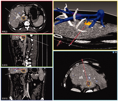
Table I. Clinical characteristics of patients who underwent microwave ablation assisted by 3D preoperative planning and conventional 2D image preoperative planning methods.
Figure 3. Images in a 45-year-old patient with a tumour in the caudate lobe. (A) Preoperative CT imaging showed that the tumour was close to the portal vein. (B) The 3D visualisation images visualised the spatial relationship of tumour and the surrounding pipe in multi-angle. (C) The 3D visualisation images visualised the shortest distance between the tumour and surrounding vessels. (D) The preoperative planning was achieved through 3D visualisation of preoperative planning system, and three needles were needed to ablate the tumour completely. (E) The contrast-enhanced CT showed complete tumour necrosis a month after microwave ablation.
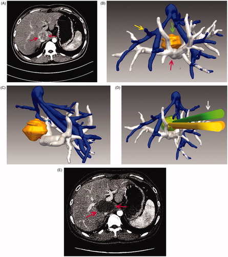
Table II. Comparison of ablation results in the 3D preoperative planning group and 2D preoperative planning group.