Figures & data
Table I. Patient and tumour characteristics.
Figure 1. Transverse MR in a 19-year-old girl with Ewing’s sarcoma in the left ilium who received curative HIFU ablation. (A) The tumour margin (arrow) was shown on T2-weighted MR before HIFU ablation. (B) The tumour (arrow) showed rich enhancement on T1-weighted contrast-enhanced MR before HIFU ablation. (C) One month after HIFU ablation, the ablation area (arrow) enveloped the entire tumour, which showed no enhancement on T1-weighted contrast-enhanced MR. (D) One year after HIFU ablation no enhancement was found in the ablation area (arrow) on T1-weighted contrast-enhanced MR.
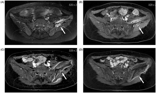
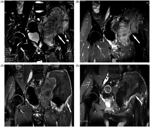
Figure 2. Coronal MR in a 15-year-old boy with osteosarcoma in the left ilium who received palliative HIFU ablation. (A) T2-weighted MR of the tumour (arrow) before HIFU ablation. (B) The tumour showed evident enhancement on T1-weighted contrast-enhanced MR before HIFU ablation. (C) After one HIFU ablation session, the ablation area showed no enhancement (white arrow), the residual tumour (black arrow) still had enhancement. (D) After the second HIFU ablation session, almost the entire tumour showed no enhancement (arrow).