Figures & data
Figure 1. A 47-year-old woman had a recurrent lymph nodule in the right neck level 3 confirmed with ultrasound-guided biopsy. (A) Ultrasound examination revealed the nodule to be 0.29 mL in volume before microwave ablation, with inhomogenous internal echoes and microcalcification. (B) Under the guidance of ultrasound, a thyroid-dedicated cooled shaft antenna (16 gauge) was positioned in the tumour. The sonogram obtained during treatment shows a typical hyperechoic region (arrow) surrounding antenna. (C) A transverse ultrasound image of the treated lymph node immediately after treatment shows increased internal echogenicity (arrow). The echogenic area (1.06 mL) was larger than the initial lymph node, suggesting the ablation zone includes surrounding normal tissue. (D) A transverse ultrasound image 3 months after treatment shows the decreased size of the ablated lymph node (arrow). (E) The transverse ultrasound image 12 months after treatment shows the ablated lymph node remaining as small scar-like tumour (arrow).
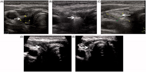
Figure 2. A 62-year-old woman with previous thyroidectomy for papillary carcinoma developed two recurrent lymph nodules in the left neck, level IV. The patient refused surgery. (A) Transverse greyscale ultrasound imaging prior to ablation revealed two rounded hypoechoic recurrent masses (measured volume = 0.10 mL, 1.17 mL, arrow) in the left neck, level IV. (B) A follow-up ultrasonogram 1 month after the ablation shows that the larger one has decreased in volume (0.37 mL, arrow), and we cannot find any evidence of the smaller one at the previous site.
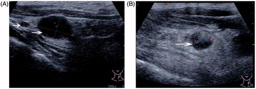
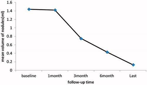
Figure 3. The mean volume change of all neck recurrences of papillary thyroid carcinoma after ultrasound-guided percutaneous microwave ablation.