Figures & data
Figure 1. Horizontal sections of MRI image showing lesion areas in persons with brain injury. The person with right-brain injury (top) and the person with left-brain injury (bottom) showed unilateral reductions in signal in the area of the posterior insula (white circles). R denotes right.
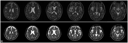
Figure 2. Schematic of the experimental design. Left panel displays a tilting response to the anodal direction in the sitting position. Participants were instructed to adjust their posture to their subjective vertical on their own accord with a notice alarm. After the notice alarm, 1.2 mA of direct current (trapezoidal pulse) was delivered to the mastoid processes via a right anode and a left cathode (right top panel). The displacement of the mean head positions during the last 3 seconds of the 5-second constant current period (grey bold line) from the time of GVS onset in each trial was analysed. Raw data from a representative healthy participant are shown (right bottom panel). Each condition consisted of 10 trials (lines) and lines show head tilting responses to the anodal direction during GVS.
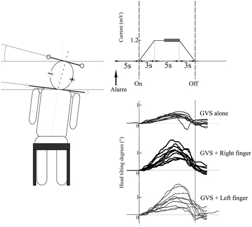
Figure 3. Mean tilting responses induced by GVS alone and GVS with finger touch in healthy participants (n = 11). The right anode stimulations with finger touch (top) induced greater tilting responses than those of the left anode (bottom). In the right anode stimulation condition, GVS with right finger touch elicited greater responses than GVS without finger touch. Error bars represent standard error. Significant differences between conditions within each anode stimulation type are shown using asterisks. *p < 0.05.
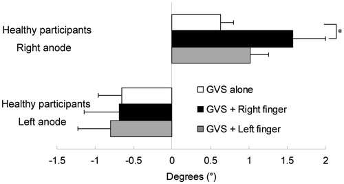
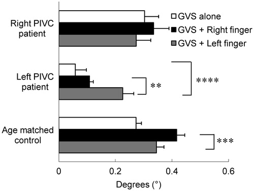
Figure 4. Tilting responses in persons with unilateral posterior insular lesion and age-matched control participants. The person with right-brain injury showed no differences in tilting responses between conditions. In the person with left-brain injury, finger touches enhanced tilting responses during GVS. In age-matched healthy participants, the right finger touch significantly modulated tilting responses. Error bars represent standard error. Only significant differences between conditions for each participant are shown using asterisks. **p < 0.01, ***p < 0.005, ****p < 0.001.