Figures & data
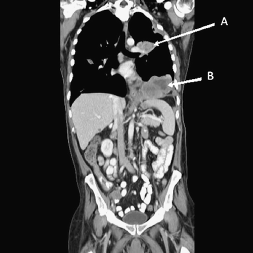
Figure 1. CT scan performed on 2/26/08 prior to sunitinib therapy. The pleural (A) and diaphragmatic (B) tumors measured 89 mm and 105.5 mm, respectively, in the vertical dimension.
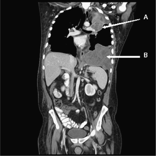
Figure 2. Histological analysis of the primary adamantinoma tumor. (A) Cellular and myxoid areas of the tumor. Scattered thick-walled blood vessels were noted (arrow). Hematoxylin and eosin; original magnification, ×200. (B) Hyalinized (asterisks) and cellular (arrow) areas of the tumor. Hematoxylin and eosin; original magnification, ×100. (C) Nuclear features of the tumor. Occasional mitotic figures (thin arrow) and focal necrosis (thick arrow) were noted. Hematoxylin and eosin; original magnification, ×400. (D) Focal positive immunostaining of the tumor for CD117 (c-kit). CD117 immunostain; original magnification, ×200. (E) Focal positive immunostaining of the tumor for platelet-derived growth factor receptor beta (PDGFR-beta). PDGFR-beta immunostain; original magnification, ×400. (F) Focal positive immunostaining of the tumor for vascular endothelial growth factor receptor-2 (VEGFR-2). VEGFR-2 immunostain; original magnification, ×200.
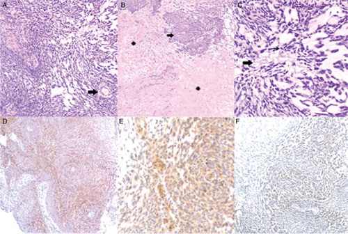
Figure 3. CT scan performed on 6/20/2008 after 4 months of sunitinib therapy. The pleural (A) and diaphragmatic (B) tumors measured 31.1mm and 57.1mm, respectively, in the vertical dimension.