Figures & data
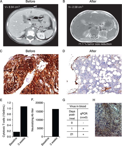
Figure 1. A chemotherapy resistant case of neuroblastoma was treated with oncolytic adenovirus Ad5/3-Cox2L-D24. A) Computer tomography of primary tumor before oncolytic virus treatment. B) Magnetic resonance imaging one month after treatment shows moderate decrease of the primary tumor and complete regression of the lymph nodes. C) Before treatment, bone marrow was almost completely infiltrated by neuroblastoma cells (CD56 staining, brown, 10x), (monoclonal mouse anti-SCLC (CD56, N-CAM), Clone 123C3, Invitrogen, CA, USA). D) Few neuroblastoma cells in bone marrow three months after oncolytic virus treatment (10x). E) Oncolytic virus treatment increased the number of CD3+/CD8+ cytotoxic T-cells in blood. F) Anti-adenoviral neutralizing antibodies were induced. G) Virus was detected in blood for three weeks after treatment. H) Immunohistochemistry indicated intermediate Cox2 expression (brown, 20x) in tumor cells (monoclonal mouse anti-human Cox-2, Clone CX229, Cayman Chemical, MI, USA).