Figures & data
Figure 1. (A) Lung carcinoma in an 81-year-old woman. Transaxial section CT scan from May 22, 2008 obtained at the level of the right pulmonary artery shows a low-density mass adjacent to the right hilus (circle). (B) Image at the same level at ten month follow-up March 2, 2009 shows progress of the mass (circle) with atelectasis ventrally. Note the cavitary air bubble within the mass (arrow). (C) 18F-FDG-PET scan from April 2, 2009 with a pathological uptake of 18F-FDG in the periphery of the tumor (arrow), but not in the central area of necrosis or in the anterior obstructive atelectasis.
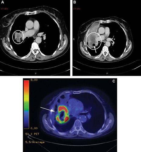
Figure 2. (A) Transverse CT image from July 3, 2008 in an 84-year-old woman with a tumor in the left upper lobe (lower picture; circle) shows soft tissue mass (upper picture; circle) between the left pulmonary artery and aorta descendens. (B) At three months after the therapy start, January 18, 2009, the primary tumor in the left upper lobe is slightly larger in volume but more confinable (lower picture; circle), whereas the lesion close to aorta descendens has become larger and necrotic (upper picture; circle). (C) 18F-FDG-PET scan from April 9, 2009 with a pathological uptake of 18F-FDG in the periphery of the expansion in front of descending aortae, but a low metabolic activity in the center of the lesion. No pathological uptake was found in the primary tumor in the left upper lobe.
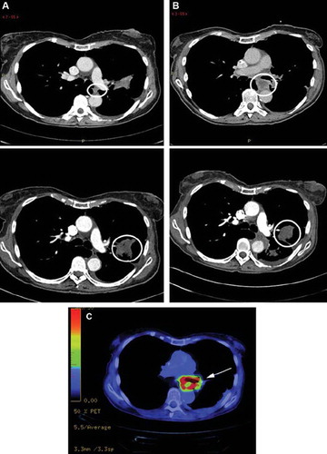
Figure 3. (A) Lung carcinoma in a 66-year-old man. Transaxial section CT scan from January 7, 2009 shows a primary tumor in the left upper lobe (circle) without any known metastasis. (B) A CT scan at the end of the study, September 23, 2009, demonstrates stable disease without any additional changes compared to previous CT scan.
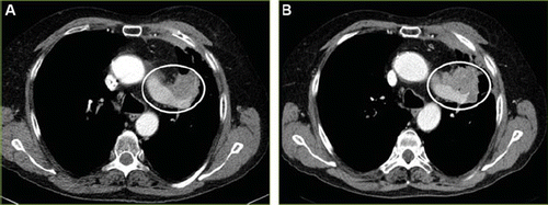
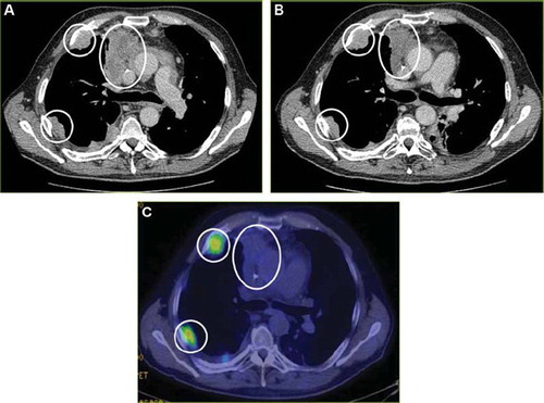
Figure 4. (A) Transverse CT image from March 17, 2009 in a 59-year-old man shows a primary tumor in the right upper lobe, as well as two metastases in the right thoracic wall (circles), but without any further metastasis. (B) A CT scan after four months of study treatment, July 21, 2009, demonstrates stable disease without any additional lesions. (C) 18F-FDG-PET scan from July 15, 2009 shows no uptake of 18F-FDG in the primary tumor and with a modest uptake in the metastases of the thoracic wall (circles).