Figures & data
Figure 1. The dose distribution and the MRIs after proton therapy: Proton beams were delivered via 3 ports (a) and the bilateral parotid glands (b), mandibular ramus (c), and cervical spine (d) in the treatment field showed retardation.
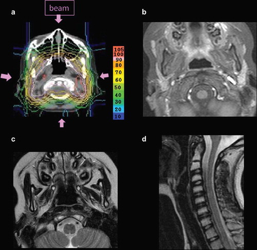
Figure 2. A salivary scintigram of 4.5 years after proton therapy. (a): The accumulation in the left parotid gland was almost normal (arrow), but reduced in the right. Salivary secretion in the oral cavity was also shown. (b): The graph shows time-dependent change of salivary flow of each salivary gland; ROI 1–5 shows right and left parotid gland, right and left submandibular gland, and control, respectively. After 20 minutes from start of the study, patient licked tartaric acid for taste stimulation. Salivary flow was observed in both parotid glands after taste stimulation, while right salivary function was depressed. Both submandibular glands showed hypoaccumulation and loss of response to taste stimulation.
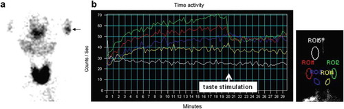
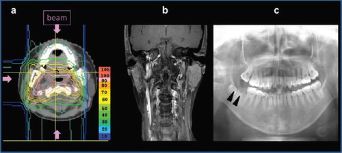
Figure 3. Proton beams were delivered via 3 ports (a). The right parotid gland (b) and the right lower back teeth (Δ:c) in the treatment field showed atrophy on the MRI and the dental panoramic radiography.