Figures & data
Figure 1. Effects of TAM on MCF-7 cell viability and apoptosis. (A) MCF-7 cells were treated with different concentrations of TAM for 24 h and 48 h. Cell growth inhibition was assessed by the MTT assay. Points represent means ± SD. Sigmoidal dose response curves were derived from fitting the data to a non-linear regression program (Graph Pad Prism). (B) MCF-7 cells were treated with different concentrations of TAM for the indicated times. The changes in cell cycle phase distribution were assessed using flow cytometry with propidium iodide (PI) staining.
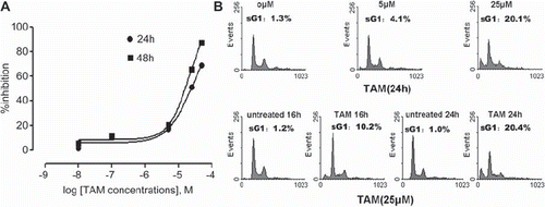
Figure 2. Role of c-Src, ERK1/2 , and AKT during TAM-induced apoptosis. (A) The cells were treated with 25μM TAM for the indicated times. The total protein was isolated for measuring phospho-Src, c-Src and β-tubulin levels by Western blotting. (B) The cells were treated with 25μM TAM for the indicated times. The total proteins were isolated for measuring phospho-ERK, ERK, phospho-AKT, AKT and β-tubulin levels by Western blotting. (C) The cells were pre-treated with 10 μM of PP2, 20 μM of PD98059, or 10 μM LY294002 for 1 h, and then treated with 25 μM TAM for 24 h. The changes in cell cycle phase distribution were assessed by flow cytometry analysis. Data are means ± SD of three independent experiments (#p < 0.05 compared to the untreated group cells; *p < 0.05 compared to cells treated with only TAM).
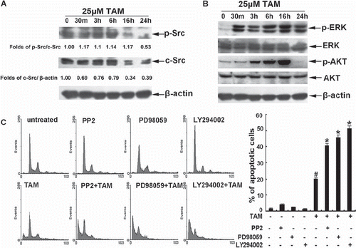
Figure 3. Effects of c-Cbl on sensitivity of the cells to TAM. (A) Cells were untreated, or treated with 25 μM TAM for 30m, 3 h, 6 h, 16 h or 24 h. The expression of c-Cbl proteins was analyzed by Western blotting. (B) Cells were transiently transfected with plasmids encoding Vec, c-Cbl or 70Z/Cbl for 48 h. The cells were then untreated or treated with 25 μM TAM for 24 h, then the changes in cell cycle phase distribution were assessed by flow cytometric analysis. The final results are summarized in the bar graphs. (C) Data are means ± SD of three independent experiments (*p < 0.05 compared to the cells in the Vec group; #p < 0.05 compared to the cells in the Vec group).
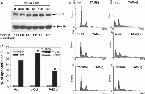
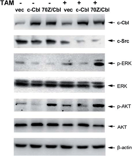
Figure 4. Effect of c-Cbl on the survival signals. Cells were transiently transfected with plasmids encoding Vec, c-Cbl or 70Z/Cbl, and then the cells were untreated, or treated with 25 μM TAM for 6 h. Then, the changes of c-Src, and the activities of ERK and AKT were analyzed by Western blotting.