Figures & data
Figure 1. Analysis of SA-sCD40L fusion protein by 12% SDS-PAGE and Western blotting. Lane 1: Protein molecular weight marker; Lane 2: Un-induced control; Lane 3: IPTG-induced expression; Lane 4: Inclusion body; Lane 5: After purification through Ni-NTA column; Lane 6: Refolded SA-sCD40L; Lane 7: Identification with hamster anti-mouse CD40L antibody.
Figure 2. Bi-functional activity of SA-sCD40L. The display of SA-sCD40L on the surface of biotinylated MB49 cells was examined by flow cytometric analysis with unbiotinylated MB49 cells as a control (Figure 2a). Indicated concentrations of SAsCD40L were incubated with 1 × 10⁵ B cells with recombinant IL-4 (10ng/ml) per well in 96-well plates. After incubation for 72 hours, cell proliferation was measured by MTT assay (Figure 2b). Furthermore, proliferation through induction of SA-sCD40L fusion protein was inhibited when anti-sCD40L antibody was added into the culture (Figure 2c).
Figure 3. Persistence of SA-sCD40L on the biotinylated mucosal surface of bladder wall. The SA-sCD40L-treated mice were killed to obtain their bladders on day 2, 4 and 6 day after intravesical instillation. Sections of 10 μm were cut and stained anti-mouse sCD40L antibody. The unbiotinylated bladder was used as a negative control.
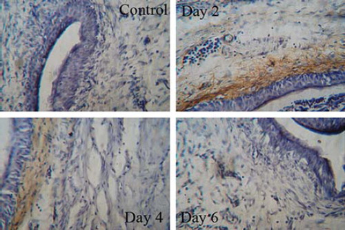
Figure 4. Immunohistochemical analysis for tumor-infiltrating lymphocytes. Bladders were removed from mice 7 days after last intravesical therapy and snap-frozen. Sections of 10 μm were cut and stained with rat-anti-mouse CD4+ or CD8+ antibody. After detection with anti-rat IgG-HRP and development by DAB display liquid kit, sections were counterstained with hematoxylin.
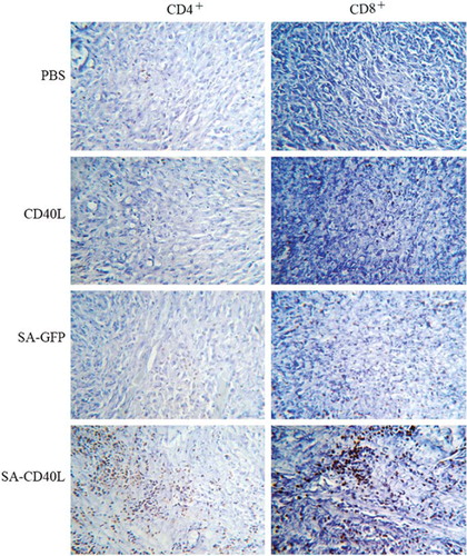
Figure 5. The therapeutic effect of SA-sCD40L fusion protein on the mouse orthotopic model of MB49 superficial bladder cancer. Mice were instilled with PBS, sCD40L, SA-GFP or SA-sCD40L one day after MB49 cells implantation. Each group had 10 mice. The survival curves were significantly different between the SA-CD40L group and the sCD40L group (p < 0.001), and between the SA-CD40L group and SA-GFP-treated group (p = 0.041).
Table I. Infiltration of CD4+ T and CD8+ T cells into the tumor tissue.