28,255
Views
44
CrossRef citations to date
0
Altmetric
Research Article

Is there a common SUV threshold in oncological FDG PET/CT, at least for some common indications? A retrospective study

, , &
Pages 670-677 | Received 04 Jun 2010, Accepted 22 Dec 2010, Published online: 19 Jan 2011

Figures & data

Table I. Summary of the distribution of benign and malignant lesions in the four subgroups. Data are numbers (%) of patients and on a patient-by-patient basis unless otherwise indicated.

Figure 1. Box-and-whisker plots of SUVmax of SPN, a: mediastinal LNs, b: cervival LNs, c: and adrenal glands, d: Closed dots represent mild outliers. The frequency of extreme outliers shown as open dots is seen only in SPNs and mediastinal LNs and is absent in cervical LNs and adrenal glands. Moreover, SUVmax between benign and malignant lesions of SNPs and mediastinal LNs overlap more than in those of cervical LNs and adrenal glands. As a result, SUVmax thresholds are positively skewed for SPNs and mediastinal LNs. Data are on a patient-by-patient basis except the adrenal glands which are on a lesion-by-lesion basis.
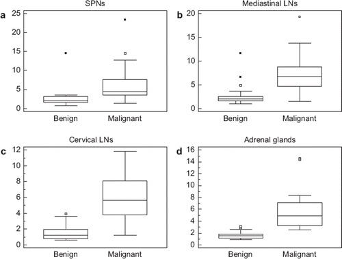
Figure 1. Box-and-whisker plots of SUVmax of SPN, a: mediastinal LNs, b: cervival LNs, c: and adrenal glands, d: Closed dots represent mild outliers. The frequency of extreme outliers shown as open dots is seen only in SPNs and mediastinal LNs and is absent in cervical LNs and adrenal glands. Moreover, SUVmax between benign and malignant lesions of SNPs and mediastinal LNs overlap more than in those of cervical LNs and adrenal glands. As a result, SUVmax thresholds are positively skewed for SPNs and mediastinal LNs. Data are on a patient-by-patient basis except the adrenal glands which are on a lesion-by-lesion basis.

Table II. Summary of lesion size and SUVmax characteristics in the four subgroups. Data for lesion size and SUVmax are median (minimum-maximum) and on a patient-by-patient basis unless otherwise indicated.

Figure 2. A 71-year-old man was admitted with delirium and fever. An MRI of the brain with and without contrast showed subacute ischemic of the left posterior frontal and parietal lobes. Blood cultures, lumbar puncture and bronchoalveolar lavage were negative for infectious pathogens. Diagnostic CT of the chest showed a right lower lung lobe nodule and an enlarged right tracheobronchial lymph node for which the FDG PET/CT was ordered for further evaluation. Axial PET (a), CT (b) and fused PET/CT (c) images demonstrated a spiculated 2.5 × 1.2 cm right lower lung lobe nodule with prominent FDG uptake (SUVmax 3.8), short arrow. This lesion (short arrow) was seen in the maximum-intensity-projection (MIP) image (d), which also showed a hypermetabolic focus in the right tracheobronchial region (long arrow). Axial PET (e), CT (f) and fused PET/CT (g) demonstrated an intensely hypermetabolic (SUVmax 4.2) right tracheobronchial lymph node measuring 1.7 × 1.5 cm in size (long arrow). The PET/CT findings were suspicious for lung primary with mediastinal metastasis. Subsequent core biopsy of the right lung nodule revealed granulomatous disease with abundant yeasts. Following antifungal treatment, the patient was afebrile on 3-month follow-up, and the right lung nodule and mediastinal lymph node resolved on chest CT.
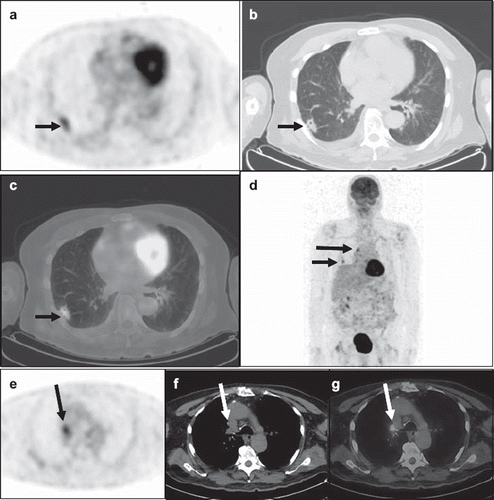
Figure 2. A 71-year-old man was admitted with delirium and fever. An MRI of the brain with and without contrast showed subacute ischemic of the left posterior frontal and parietal lobes. Blood cultures, lumbar puncture and bronchoalveolar lavage were negative for infectious pathogens. Diagnostic CT of the chest showed a right lower lung lobe nodule and an enlarged right tracheobronchial lymph node for which the FDG PET/CT was ordered for further evaluation. Axial PET (a), CT (b) and fused PET/CT (c) images demonstrated a spiculated 2.5 × 1.2 cm right lower lung lobe nodule with prominent FDG uptake (SUVmax 3.8), short arrow. This lesion (short arrow) was seen in the maximum-intensity-projection (MIP) image (d), which also showed a hypermetabolic focus in the right tracheobronchial region (long arrow). Axial PET (e), CT (f) and fused PET/CT (g) demonstrated an intensely hypermetabolic (SUVmax 4.2) right tracheobronchial lymph node measuring 1.7 × 1.5 cm in size (long arrow). The PET/CT findings were suspicious for lung primary with mediastinal metastasis. Subsequent core biopsy of the right lung nodule revealed granulomatous disease with abundant yeasts. Following antifungal treatment, the patient was afebrile on 3-month follow-up, and the right lung nodule and mediastinal lymph node resolved on chest CT.

Table III. Summary of SUVmax thresholds and corresponding sensitivities, specificities, positive predictive values (PPV), negative predictive values (NPV) as well as AUC for the four subgroups. Data are on a patient-by-patient basis unless otherwise indicated.

Reprints and Corporate Permissions

Please note: Selecting permissions does not provide access to the full text of the article, please see our help page How do I view content?

To request a reprint or corporate permissions for this article, please click on the relevant link below:

Academic Permissions

Please note: Selecting permissions does not provide access to the full text of the article, please see our help page How do I view content?

Obtain permissions instantly via Rightslink by clicking on the button below:

If you are unable to obtain permissions via Rightslink, please complete and submit this Permissions form. For more information, please visit our Permissions help page.