Figures & data
Figure 1. Histopathology of DMBA and MAT B III tumors. Paraffin sections stained with hematoxylin/eosin. Bars: A and B 100 μm; C and D 50 μm. A. Light micrograph showing a DMBA tumor. Cords of tumor cells form gland-like structures, which are separated by strands of dense connective tissue. Note the distinct, thick capsule. Htx/E. B. Light micrograph of a MAT B III tumor, showing low differentiation of the densely packed tumor cells, absence of organized structures, scanty extracellular matrix and irregular infiltration by tumor cells in the surrounding connective tissue. C. Higher magnification of a DMBA tumor, showing the differentiated glandular appearance of the tumor. D. Higher magnification of a MAT B III tumor, showing the densely packed pleomorphic tumor cells. Occasional mitotic figures are seen.
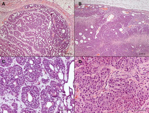
Figure 2. IFP measurement setup. A. An anaesthetized Fisher rat with two bulging ellipsoid subcutaneous MAT B III tumors. A FOPT is inserted in each tumor, guided by a Venflon™ tube. Plastic clay applied to prevent leakage. B. A FOPT with a sensor in its distal (left) end is shown resting on a finger tip. Note the small dimensions. C. A 0.5 mm space, filled with fluid in situ, is located between the orifice of the PTFE tube and the sensor (dark), enclosed by a protective PTFE tube. D. A side view of a FOPT showing to the left the sensor as a dark spot, inserted in a protecting PTFE tube. This construction prevents direct load on the sensor by the tissue, which may cause faulty recordings.
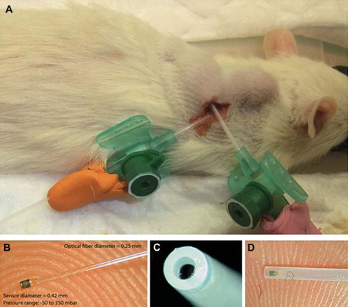
Table I. Effects of AF-16 on IFP in DMBA tumors and in MAT BIII tumor.
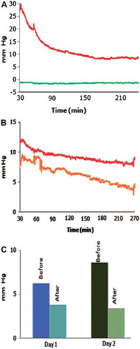
Figure 3. Effects of AF-16 on the IFP in DMBA tumors (A, B), and in MAT B III tumor (C). A. Recording of the IFP in central parts of a DMBA tumor starting 30 min after an intracapsular injection of 500 μg/kg bw of AF-16. The tumor IFP (red) decreased in 3 h to less than 40% of the pretreatment level. The IFP in adjacent normal subcutaneous connective tissue (green) remained unaffected. B. The IFP in two adjacent, but non-communicating tumors (red and yellow) decreased in parallel after intravenous injection of 500 μg/kg bw of AF-16. C. Injection of 500 μg/kg bw of AF-16 once daily in the capsule zone of a MAT B III tumor during two consecutive days reduced the IFP both days.