975
Views
11
CrossRef citations to date
0
Altmetric
Research Article

Inflammatory cells in node-negative breast cancer

, , , , , & show all
Pages 680-686 | Received 19 Sep 2011, Accepted 16 Dec 2011, Published online: 24 Jan 2012

Figures & data

Table I. Patients’ characteristics.

Table II. Descriptive statistics. Mean, median, minimal and maximal values of the different cells types presented.

Figure 1. Immunohistochemical stainings. Membraneous stainings of CD3, CD4, CD8 and FoxP3 (nuclear staining) all markers of different subgroups of T-cells. Membraneous staining of CD20 (B-cells). Cytoplasmic stainings of CD68 (macrophages) and tryptase (mast cells).

Figure 1. Immunohistochemical stainings. Membraneous stainings of CD3, CD4, CD8 and FoxP3 (nuclear staining) all markers of different subgroups of T-cells. Membraneous staining of CD20 (B-cells). Cytoplasmic stainings of CD68 (macrophages) and tryptase (mast cells).

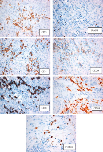
Table III. Spearman correlation. Each parameter was correlated to each other.

Table IV. Conditional logistic regression. Case-control study. End point: breast cancer death. Models adjusted for tumour size and age at diagnosis. Different cut-off values were tested in explorative analyses and cut-off at 50 percentile is presented.

Reprints and Corporate Permissions

Please note: Selecting permissions does not provide access to the full text of the article, please see our help page How do I view content?

To request a reprint or corporate permissions for this article, please click on the relevant link below:

Academic Permissions

Please note: Selecting permissions does not provide access to the full text of the article, please see our help page How do I view content?

Obtain permissions instantly via Rightslink by clicking on the button below:

If you are unable to obtain permissions via Rightslink, please complete and submit this Permissions form. For more information, please visit our Permissions help page.