Figures & data
Table I [Citation4,Citation10].
Table II. The average survival time for patients with HPCs [Citation1].
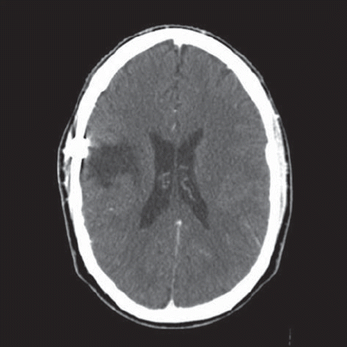
Figure 1. First CT-scan performed after i.v. contrast injection shows a right-sided frontoparietal haemorrhagic lesion (5 × 6 cm) with surrounding oedema, a right-sided temporofrontal subdural haematoma of 7 mm and a marked (12 mm) shift of the midline structures.
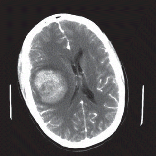
Figure 2. Highly cellular tumour tissue, displaying nuclear polymorphism and mitoses (arrows) (Haematoxylin-eosin. Bar: 100 μm).
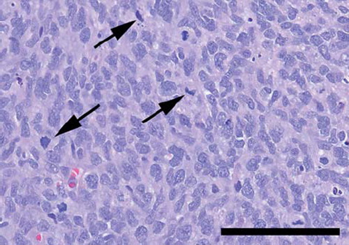
Figure 3. There were numerous necroses throughout the tumour (Haematoxylin-eosin. Bar 500 μm).
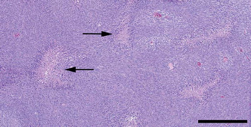
Figure 4. The tumour tissue was rich in compressed, thin-walled blood vessels. (Anti CD34 – haematoxylin. Bar: 100 μm). (b) Thin-walled sinusoidal blood vessels, forming staghorn patterns were found throughout the tumour (Anti CD34 – haematoxylin. Bar: 200 μm).
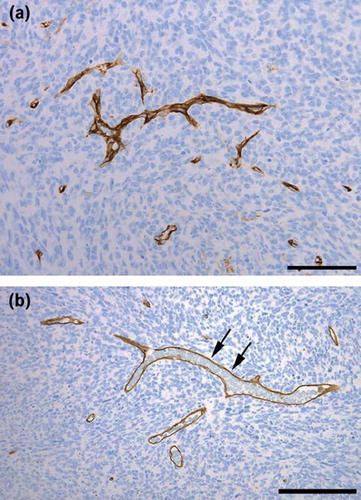
Figure 5. Five weeks postoperatively, CT-scan performed after i.v. contrast injection, showing no signs of remaining tumour and the haematoma is totally absorbed.