Figures & data
Table I. Overview of the implementation of DBCG-IMN radiotherapy guidelines, non-CT-based techniques for IMN radiotherapy and the transition from simulator-based to 3D-CT-guided dose planning in the DBCG radiotherapy departments.
Figure 1. Field arrangements in the five radiotherapy techniques. Left: A: Left-sided two field arrangement in post-mastectomy patients (PM Left), B: Right-sided three field arrangement in post-mastectomy patients (PM Right), C: Left-sided standard tangents in post-lumpectomy patients (PL Left), D: Right-sided wide tangents in post-lumpectomy patients (PL Right T) and E: Right-sided electron field combined with tangential fields in post-lumpectomy patients (PL Right E + T). Black: Shielded areas. Light grey: Photon fields. Dark grey: Electron fields. Right: Axial slices: Dashed Lines: Electron field borders, Solid lines: Photon field borders. Dotted lines: Field axis.
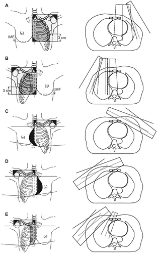
Figure 2. Internal mammary node dose coverage in the different techniques. Horizontal lines: Mean. Filled circles: individual patient dose coverage estimates.
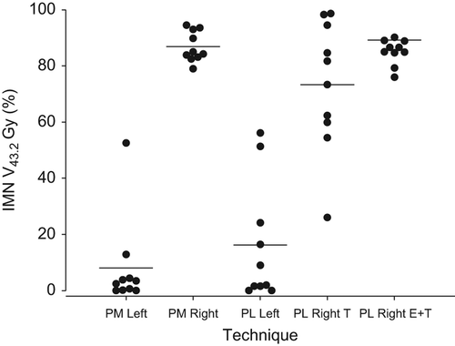
Figure 3. Internal mammary node point doses at a point within the internal mammary node clinical target volume contour in the third intercostal space.
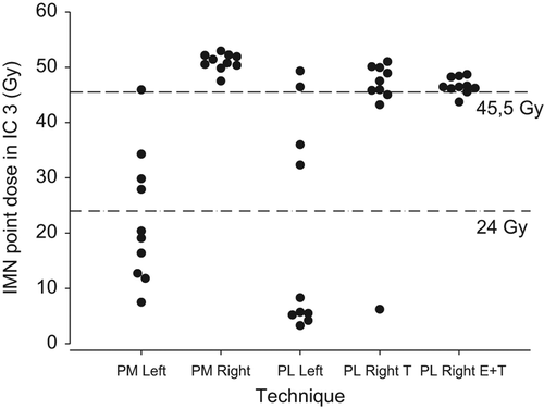
Table II. Summary of dose coverage estimates for internal mammary lymph nodes and organs at risk in the five standard DBCG techniques.