Figures & data
Table I. Results from the comparison of the automated segmentation and the manual reference delineation.
Figure 1. Examples of the prostate segmentation in the axial plane (left) and the sagittal plane (right) from three patients. Blue: Manual reference segmentation, Green: Automated segmentation, Red: Average model. The DSC for the three segmentations was 0.94 (top), 0.79 (middle), 0.89 (bottom).
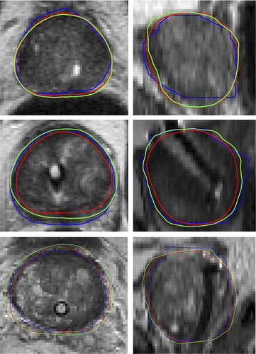
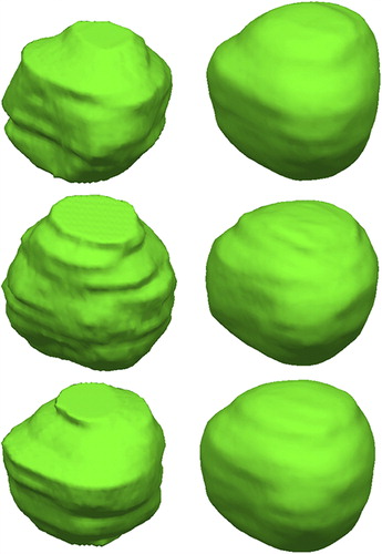
Figure 2. Examples of the prostate segmentation. Left: The reference manual segmentation. Right: Automated 3D segmentation.
Supplemental material