
Figures & data
Figure 1. (a) The external marker block position for patient group 1 and 2. Field 1 (F1), field 2 (F2) and the u direction are indicated. (b) A schematic drawing of the marker block motion before and during a DIBH, indicating the gating level and gating window. (c) An example of a portal image of field 2. The relative deviation of the chest wall was found by comparison of the pixel intensities in three regions of interest as shown. The u (and v) directions are indicated by arrows.
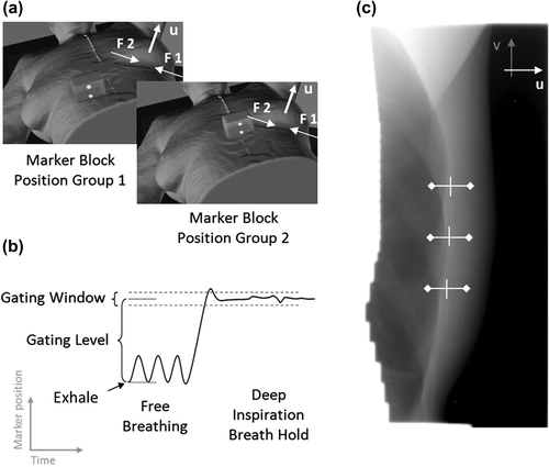
Table I. Patient characteristics and treatment information for patient groups 1 and 2.
Figure 2. Cumulative frequencies of (a) absolute inter-fraction setup error, (b) absolute inter-field shift and (c) intra-field motion range for group 1 and 2.
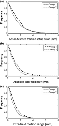
Figure 3. The time resolved setup errors of the chest wall for (a) all imaged fractions and (b) the first treatment fraction for a typical patient. For the first fraction, the figure also shows (c) the external marker block position and (d) the resulting gate signal, which is in “enable state” when the marker block is inside the gating window [- - - -].
![Figure 3. The time resolved setup errors of the chest wall for (a) all imaged fractions and (b) the first treatment fraction for a typical patient. For the first fraction, the figure also shows (c) the external marker block position and (d) the resulting gate signal, which is in “enable state” when the marker block is inside the gating window [- - - -].](/cms/asset/85f56072-76cd-4630-9af5-1ca69dfbbe5c/ionc_a_1045625_f0003_b.gif)
Figure 4. Box plots of the patient individual (a) inter-fraction setup error (b) inter-field shift and (c) intra-field motion range. The box indicates the 25th and 75th percentiles, while the extending narrow bars indicate the most extreme data points that are not considered outliers [o].
![Figure 4. Box plots of the patient individual (a) inter-fraction setup error (b) inter-field shift and (c) intra-field motion range. The box indicates the 25th and 75th percentiles, while the extending narrow bars indicate the most extreme data points that are not considered outliers [o].](/cms/asset/6271f3df-af3d-4afe-97bc-e13a3d278818/ionc_a_1045625_f0004_b.gif)
Figure 5. Three examples of internal chest wall setup error and external marker block position during fractions with large inter-field shifts (a, c) and intra-field motion (b, c). The gating window (---) and resulting binary beam-enable signal (bottom graph) are indicated for each case.
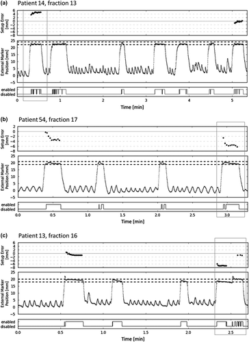
Table II. Gating and imaging characteristics for patient groups 1 and 2.
Table III. The inter-fraction setup error (fields 1 + 2), inter-field shift, and intra-field motion for patient group 1 and 2, individually and combined.