Figures & data
Table 1. The regions of different structural appearances in the individual tendons, displayed as percentage of total section area
Table 2. The labeling of COMP and type I and III collagens is displayed in the different regions of the tendon in each individual, as immunopositive staining, graded as 0, 1, 2, and 3, where 0 represents no staining and 3 represents a marked staining
Figure 1. Microscopic longitudinal section (H&E stained) of a normal tendon (L). Note the aligned fiber bundles with tenocytes in parallel rows and thin endotenon (arrows). L refers to tendon ID (see Tables 1 and 2). Bar = 100 μm.
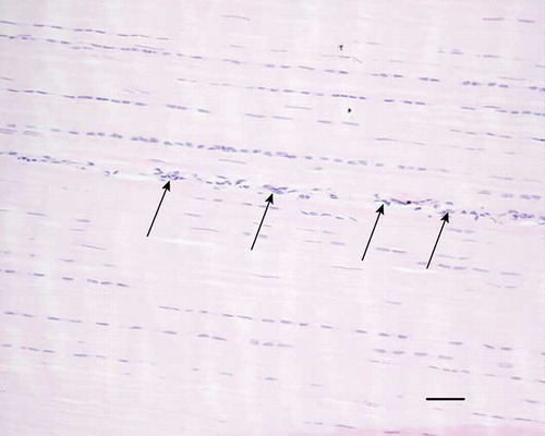
Figure 2. Microscopic longitudinal section (H&E stained) of an injured tendon (C) displaying an organized fibroblastic region. There is a high cellularity with fibroblastic cells and fibers in longitudinal parallel arrangement. Vessels are present between fibers (arrows). C refers to tendon ID (see Tables 1 and 2). Bar = 200 μm.
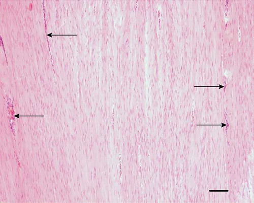
Figure 3. Microscopic longitudinal section (H&E stained) of an injured tendon (H) showing a disorganized region with high amount of fibroblastic cells (arrows) and many vessel-like structures (arrowheads) compatible with neovascularization. H refers to tendon ID (see Tables 1 and 2). Bar = 100 μm.
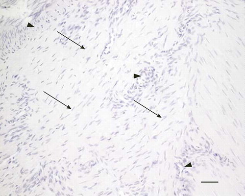
Figure 4. Microscopic longitudinal section (H&E stained) of injured tendon (A) characterized by acellular necrotic area (arrows) and hemorrhages (*). A refers to tendon ID (see Tables 1 and 2). Bar = 100 μm.
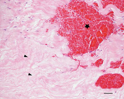
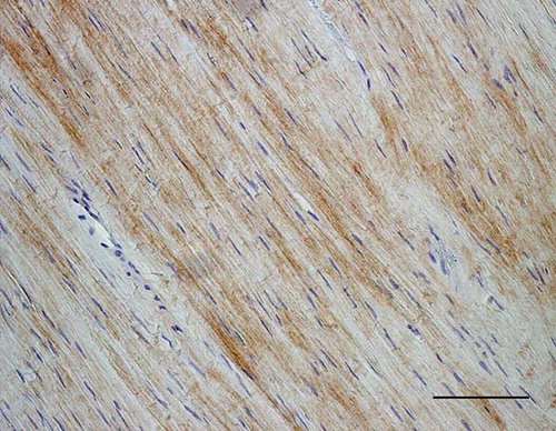
Figure 5. Microscopic longitudinal section of a normal tendon (K). Immunohistochemical localization of COMP. Note the staining in the normally aligned fibers. K refers to tendon ID (see Tables 1 and 2). Bar = 100 μm.
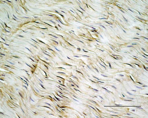
Figure 6. Microscopic longitudinal section of an injured tendon (C). A: Immunohistochemical localization of COMP in a fibroblastic organized region with a marked staining (arrows) and in endotenon (arrowheads). Bar = 100 μm. B: In situ hybridization with the expression of COMP m-RNA (dark stain) in the cytoplasm of the majority of cells (arrows) in the same region as a). C refers to tendon ID (see Tables 1 and 2). Bar = 100 μm.
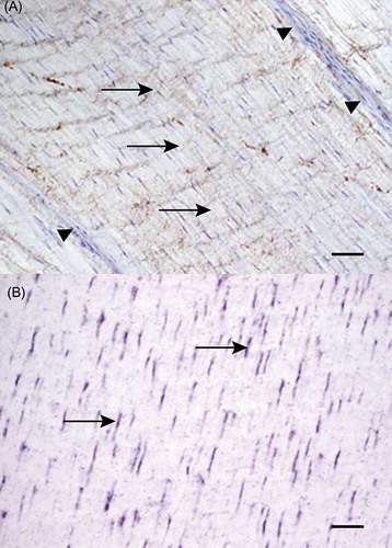
Figure 7. Microscopic longitudinal section of injured tendon (H). Immunohistochemical localization of type I collagen. Note the intense staining of the organized fibroblastic tissue, but lack of staining in the extensive endotenon (arrows). H refers to tendon ID (see Tables 1 and 2). Bar = 100 μm.
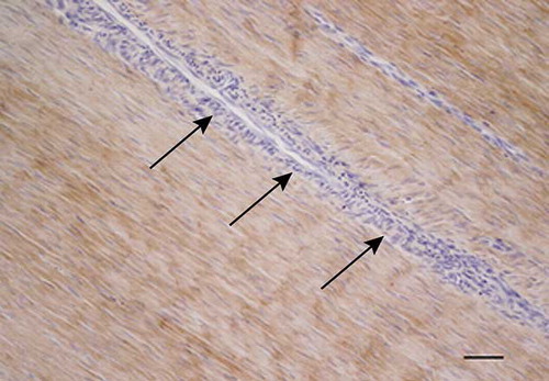
Figure 8. Microscopic longitudinal section of a normal tendon from a control (K). Immunohistochemical localization of type III collagen. A marked immunostaining is present in the endotenon (arrows), but the aligned fibers do not show any staining. K refers to tendon ID (see Tables 1 and 2). Bar = 100 μm.
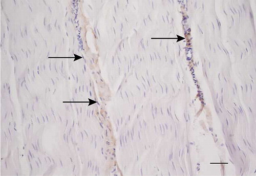
Figure 9. Microscopic longitudinal section of an injured tendon (B). Immunohistochemical localization of type III collagen. A marked immunostaining is present in an atypical, disorganized region. B refers to tendon ID (see Tables 1 and 2). Bar = 100 μm.