Figures & data
Figure 1. Time taken by the animals to enter the dark compartment of the passive avoidance apparatus during the exploration trials of passive avoidance test. The entrance latency to the dark compartment was decreased in both the groups from first to third trial, but there was a significant difference in the entrance latency time of the groups in the second and third trials. The radio-frequency electromagnetic radiation (RF-EMR)-exposed animals took more time to enter the dark compartment during the exploration trials. *P < 0.05.
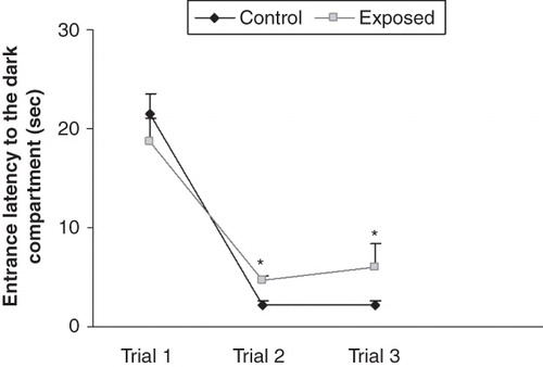
Figure 2. Effect of radio-frequency electromagnetic radiation (RF-EMR) on latency to enter the dark compartment 24 hours (A) and 48 hours (B) after the shock trial. Rats exposed to the mobile phone took significantly less time to enter the dark compartment in the memory retention test. Results are expressed as mean ± SEM. *P < 0.05.
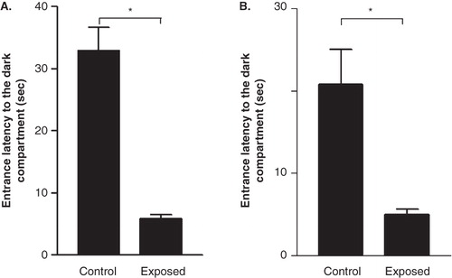
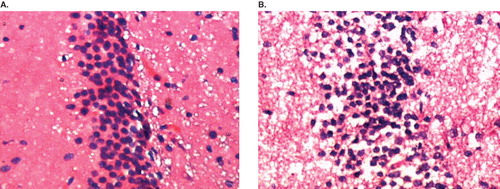
Figure 3. Representative photomicrograph of sections of hippocampal CA3 region of the brain from both control and radio-frequency electromagnetic radiation (RF-EMR)-exposed rat stained with hematoxylin and eosin. A: Control animal; row of normal nerve cells in a section of the pyramidal cell band of the hippocampus CA3 region is seen. B: Mobile phone RF-EMR-exposed rat; among the normal nerve cells, dark (deeply stained) and shrunken nerve cells are seen.