Figures & data
Winner of the Rudbeck Award, 2010 at the Medical Faculty of Uppsala University.For his pioneering research on the mechanisms of islet amyloidosis and its impact on the pathogenesis of type 2 diabetes.
Figure 1. Pancreatic section of case IsN13 with islets filled with amyloid, stained with Congo red. This material was used to purify the IAPP giving the first amino acid sequence from human islet origin. There are signs of pronounced autolysis, which did not affect the quality of the purified peptide. There was also partial exocrine atrophy which was helpful in that it made the amyloid more concentrated.
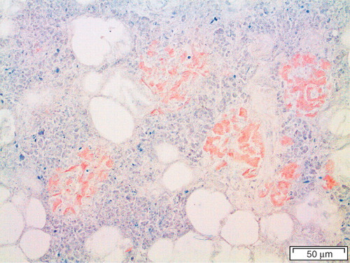
Figure 2. Pancreatic section of a common shrew (Sorex araneus), labeled with antibodies against IAPP. There is a strong reaction with the β-cells, which constitute the majority of islet cells.
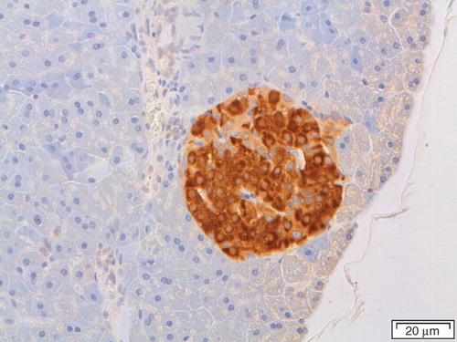
Figure 3. Pancreatic section of a degu (Octodon degus) with heavy amyloid deposits at the islet periphery, stained with Congo red.
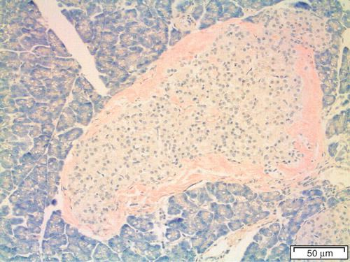
Figure 4. Human islet from a diabetic subject. Most of the islet has been converted into amyloid, but there are still cords of cells, a majority being β-cells, most probably dysfunctional. Congo red. Bar: 50 μm.
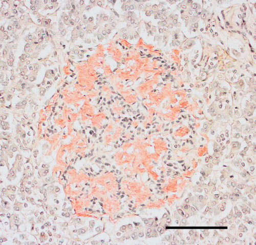
Figure 5. Section from the pancreas of a diabetic subject with severe islet infiltration of amyloid in all islets, visualized with Congo red. The section in A is seen in ordinary light, while in B polarized light with crossed polars has been used. A bright yellow birefringence is evident.
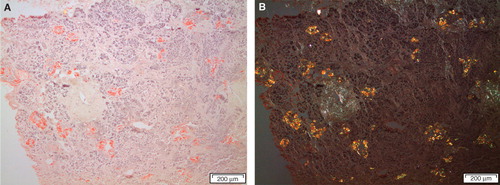
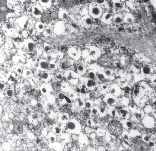
Figure 6. Electron micrograph showing a part of a β-cell from a diabetic subject. There are bundles of typical amyloid fibrils which penetrate deeply into the cells. Note that the cell is filled with characteristic insulin granules.