Figures & data
Figure 1. A: Ductal carcinoma in situ of the breast, the comedo form, with several cell layers of epithelial cells surrounding a central necrotic area. The inner cell layers, adjacent to the necrosis, show low differentiation with unorganized structures and increased nucleus-to-cytoplasm ratio (scale bar: 500 μm). B: Non-malignant mammary epithelial cells cultured in a three-dimensional differentiation-inducing assay. After 21 days of culture at normoxia (21% O2), the mammary epithelial cells (MCF-10A) differentiate into growth-arrested, organized acini structures with polarized cells surrounding a hollow lumen, resembling the in vivo mammary gland appearance. The differentiated mammary cells have a polarized expression pattern of proteins, here laminin V (green), and small compact nuclei (blue, actin in red) in a palisade structure. C: At hypoxia (1% O2) the MCF-10A mammary epithelial cells fail to arrange into organized structures and appear as cell aggregates without lumen or polarized protein localization. The hypoxic cells have larger nuclei, remain proliferative, and express markers of undifferentiated cell stage—characteristics often seen in breast carcinoma (scale bar: 20 μm).
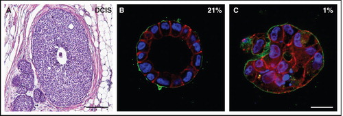
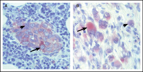
Figure 2. Human neuroblastoma specimens stained for neuron-specific enolase by immunohistochemistry. Sections of a neuroblastoma bone-marrow metastasis (A) and a ganglioneuroma specimen (B), respectively, stained for neuron-specific enolase (ENO2) expression. Note that tumor cells differ considerably in neuron-specific enolase levels, both at a more immature (panel A) and at a differentiated (panel B) stage. Arrows show enolase-positive and arrow-heads show enolase-negative tumor cells.