Figures & data
Figure 1. Lateral plain radiograph of the thoracic spine. There were mild degenerative changes in T3 to T11 vertebral bodies and discs.
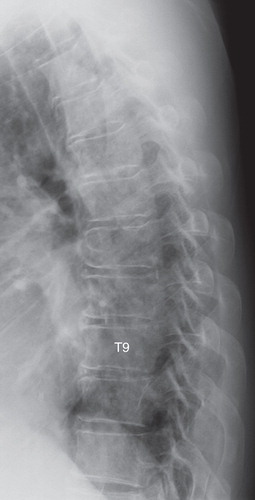
Figure 2. T2-weighted MR images of the thoracic spine. The ligamentum flavum at the right T9–10 level was thickened (arrows). A: sagittal view. B: axial view.
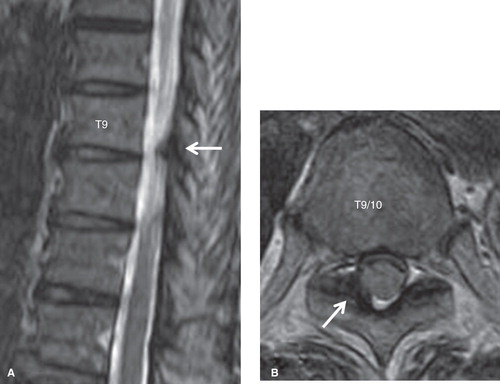
Figure 3. CT images after conventional myelography of the thoracic spine. OLF at the right T9–10 (A) and bilateral T10–11 (B) levels were observed (arrows).
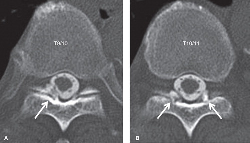
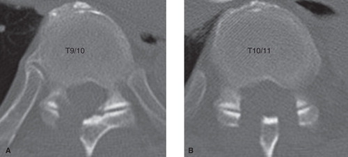
Figure 4. CT images of the thoracic spine after the operation. Sufficient decompression was accomplished at the right T9–10 (A) and bilateral T10–11 (B) levels.