Figures & data
Figure 1. Palpable purpuric rash on the left lower extremity (A and B) and back (C and D) following antibiotic therapy.
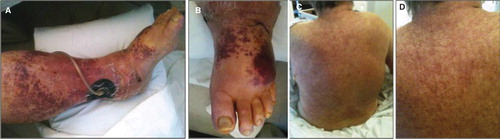
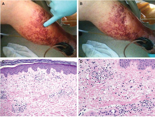
Figure 2. Demonstration of non-blanchability of the purpuric lesions (A and B). Histopathology of skin lesions from the purpuric lesions (C). The epidermis demonstrates no inflammatory changes. Within the papillary dermis there is a mild inflammatory infiltrate associated with extravasated red blood cells (× 100, H&E). The inflammatory infiltrate is composed predominately of lymphocytes with rare eosinophils (D). There are scattered extravasated red blood cells. No neutrophils are present. The vessels show no fibrinoid necrosis or fibrin thrombi (× 200, H&E).