Figures & data
Figure 1. Systemic circulatory variables: mean blood pressure, end-tidal PCO2, coronary perfusion pressure, cerebral cortical blood flow, and cardiac output. Coronary perfusion pressure is presented both for the whole experiments and for the CPR period only. Vasopressin group •; Vasopressin-adrenaline group ▪.
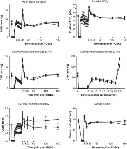
Figure 2. Systolic and mean blood pressures as related to cerebral cortical blood flow before cardiac arrest, during CPR, and immediately after restoration of spontaneous circulation. Vasopressin group •; Vasopressin-adrenaline group ▪.
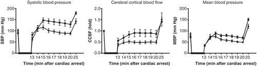
Figure 3. Systemic circulatory variables in the background study for surviving and non-surviving animals. Surviving animals ▴; non-surviving animals ▾.
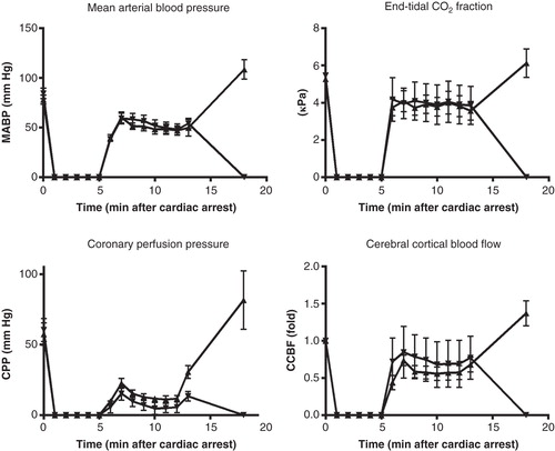
Figure 4. Tissue injury indicators: troponin I, protein S-100β, 8-iso-dihydro-PGF2α. Vasopressin group •; Vasopressin-adrenaline group ▪.
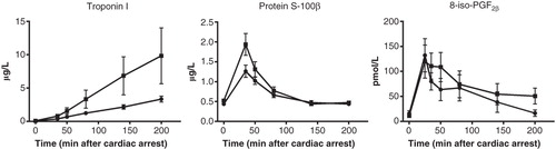
Figure 5. Tissue injury indicators for surviving animals in the background study.
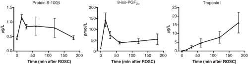
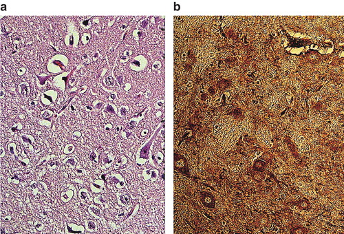
Figure 6. Leakage of albumin into the neuropil of cerebral cortex in pig brain after cardiac arrest and treatment with vasopressin and adrenaline (VA group) (a) and vasopressin alone (V group) (b). Albumin leakage in the neuropil and neurons was considerably reduced in pigs treated with vasopressin alone in comparison with the group receiving vasopressin and adrenaline. ×300.
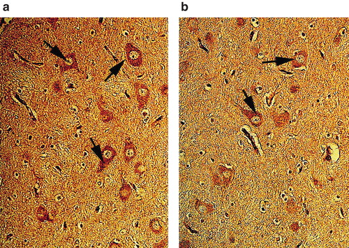
Figure 7. Nissl-stained neurons in the neuropil of cerebral cortex in pigs treated with vasopressin and adrenaline in combination (VA group) (a) or vasopressin alone (b). Vasopressin alone caused less neuronal damage, sponginess, and edema up to a certain extent in pigs after cardiac arrest, while several neurons were damaged with perineuronal edema in the group given combined treatment with vasopressin and adrenaline (VA group). ×300.
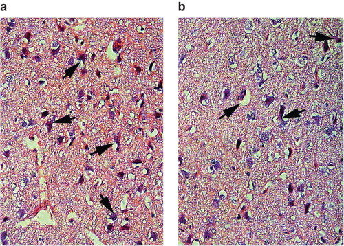
Figure 8. Box plots (median, 25th and 75th as well as 10th and 90th percentiles) of microscopical assessment of neuronal injury and albumin leakage (vasopressin group, vasopressin-adrenaline group, and background study) and correlation of these two variables for the vasopressin and vasopressin-adrenaline groups (R2 = 0.85, P < 0.0001).
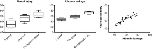
Figure 9. Neuronal damage (a) and albumin leakage (b) in the cerebral cortex of pigs treated with adrenaline only during CPR after cardiac arrest (background study). Massive neuronal damage with perineuronal edema and sponginess was evident (b). There was profound leakage of albumin in the neuropil and strong albumin-positive nerve cells as well (b). ×300.