Figures & data
Figure 1. Characteristic diffuse flame-shaped hemorrhages and edema in the posterior pole in a patient with central retinal vein occlusion.
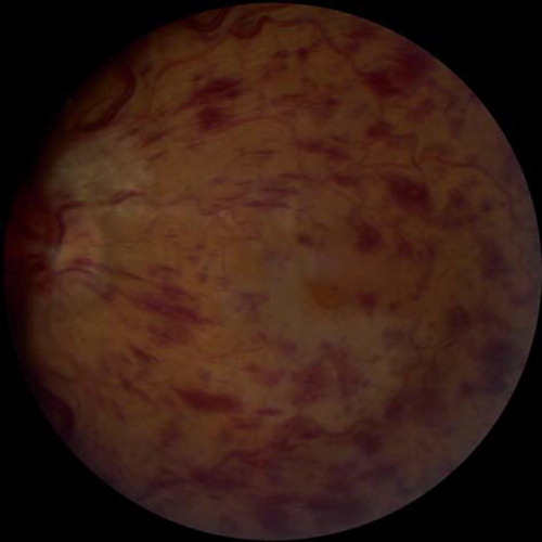
Figure 2. Fundus photograph demonstrating accumulation of lipid exudates in a patient with clinically significant diabetic macular edema.
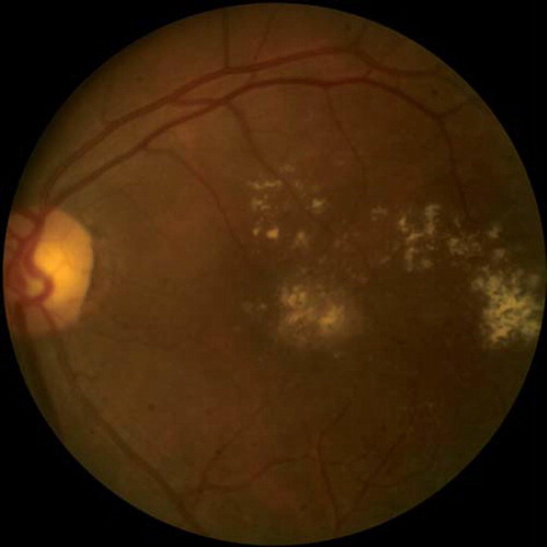
Figure 3. Fundus photograph demonstrating accumulation of lipid exudates in a patient with clinically significant diabetic macular edema.
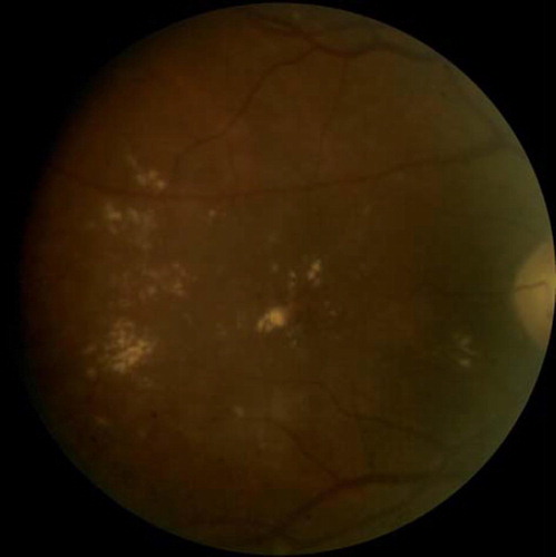
Figure 4. Tractional retinal detachment secondary to proliferative diabetic retinopathy. Note presence of neovascularization of the disc as well as elsewhere.
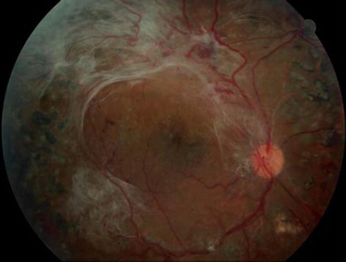
Figure 5. Well circumscribed sub-hyaloid hemorrhage in a patient with proliferative diabetic retinopathy.
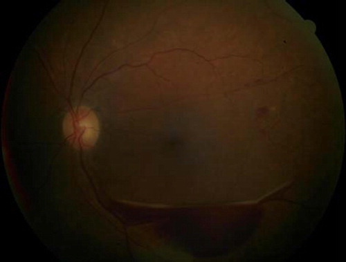
Figure 6. Bilaterally enlargement of extraocular muscles, especially in the inferior and medial rectus muscles, in a patient with thyroid eye disease.
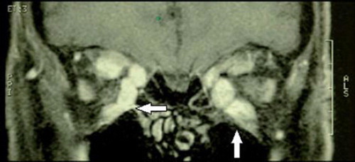
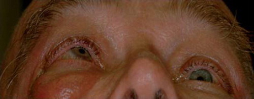
Figure 7. A patient with thyroid eye disease exhibiting prominent exophthalmos that is greater in the right eye, with significant restriction of right eye movement.
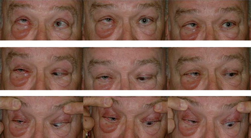
Figure 8. A patient with thyroid eye disease exhibiting prominent exophthalmos that is greater in the right eye with significant restriction of right eye movement.