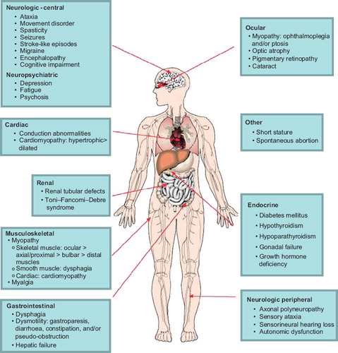
8,798
Views
113
CrossRef citations to date
0
Altmetric
Review Article

Diagnosis and treatment of mitochondrial myopathies

&
Pages 4-16 | Received 01 Aug 2011, Accepted 15 Aug 2011, Published online: 25 Aug 2011

Figures & data

Figure 1. Clinical features of mitochondrial myopathies, by organ system.

Figure 1. Clinical features of mitochondrial myopathies, by organ system.

Table I. Mitochondrial myopathy syndromes presenting with ocular myopathy.

Table II. Mitochondrial myopathy typically presenting without PEO.

Table III. Confirmatory tests for organ dysfunction in mitochondrial myopathy.

Figure 2. Abnormalities on skeletal muscle biopsy in mitochondrial myopathy. Serial sections through vastus lateralis in a patient with mitochondrial myopathy showing: (A) haematoxylin and eosin, (B) cytochrome c oxidase histochemistry (COX) (note the COX deficient fibres), (C) succinate dehydrogenase histochemistry (SDH) (note the sub-sarcolemmal accumulation of mitochondria analogous to a ragged red fibre), and (D) sequential COX-SDH histochemistry showing a mosaic COX defect as seen in patients with mtDNA disorders.

Figure 2. Abnormalities on skeletal muscle biopsy in mitochondrial myopathy. Serial sections through vastus lateralis in a patient with mitochondrial myopathy showing: (A) haematoxylin and eosin, (B) cytochrome c oxidase histochemistry (COX) (note the COX deficient fibres), (C) succinate dehydrogenase histochemistry (SDH) (note the sub-sarcolemmal accumulation of mitochondria analogous to a ragged red fibre), and (D) sequential COX-SDH histochemistry showing a mosaic COX defect as seen in patients with mtDNA disorders.

Table IV. Treatments with reported benefit in mitochondrial myopathy which may benefit from further study in blinded placebo-controlled trials.