3,440
Views
145
CrossRef citations to date
0
Altmetric
Review Article

The molecular basis of the frontotemporal lobar degeneration–amyotrophic lateral sclerosis spectrum

, &
Pages 817-828 | Received 21 Apr 2011, Accepted 07 Feb 2012, Published online: 16 Mar 2012

Figures & data

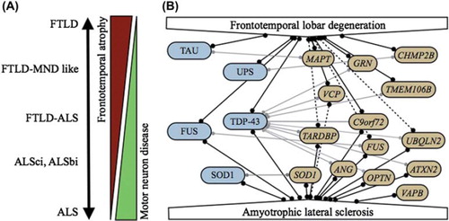
Table I. Diagnostic categories of the FTLD-ALS disease spectrum.

Figure 1. A: FTLD and ALS form a clinical disease continuum. B: Molecular relationships between FTLD and ALS. Pathological proteins are indicated in blue, causal genes in yellow. Full lines indicate strong correlations; dotted lines represent putative correlations. Correlations between genes and pathological proteins are indicated by light gray lines.

Figure 1. A: FTLD and ALS form a clinical disease continuum. B: Molecular relationships between FTLD and ALS. Pathological proteins are indicated in blue, causal genes in yellow. Full lines indicate strong correlations; dotted lines represent putative correlations. Correlations between genes and pathological proteins are indicated by light gray lines.

Figure 2. TDP-43 proteinopathy in FTLD. Ubiquitin and TDP-43 immunohistochemistry of sections from the superficial frontal cortex (FCx) and temporal cortex (TCx) of a patient with a VCP mutation (Citation176). Frequent intranuclear inclusions (arrow-heads) are seen that stain positive for ubiquitin and TDP-43.

Figure 2. TDP-43 proteinopathy in FTLD. Ubiquitin and TDP-43 immunohistochemistry of sections from the superficial frontal cortex (FCx) and temporal cortex (TCx) of a patient with a VCP mutation (Citation176). Frequent intranuclear inclusions (arrow-heads) are seen that stain positive for ubiquitin and TDP-43.

Table II. Genes associated with FTLD and ALS.

Table III. Characteristics of chromosome 9p-linked FTLD-ALS families.