Figures & data
Figure 1. Two-dimensional gel map of Aplysia abdominal ganglia proteins with statistically significant different levels after serotonin treatment. Aplysia abdominal ganglia proteins were extracted, and 700 μg were applied on an immobilized pH 3–10 non-linear gradient strip, followed by 9%–16% linear gradient polyacrylamide gel. Gels were stained with Coomassie Blue, spots were analyzed, and proteins were assigned using MASCOT software (Citation14). UniProtKB accession numbers for protein identification are given.
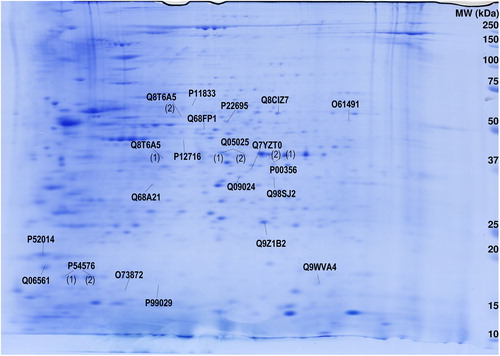
Figure 2. Verification of quantitative results from 2-DE by Western blotting in abdominal ganglia. Protein level changes after serotonin treatment in abdominal ganglia of ADF detected by (A) 2-DE and (B) Western blotting; DAZAP-1 detected by (C) 2-DE and (D) Western blotting; Flotillin-1 detected by (E) 2-DE and (F) Western blotting. Inserts in B, D, and F depict representative images of original Western blots of target proteins and β-tubulin loading control, respectively. All data display optical densities as mean ± SEM. *P < 0.05; **P < 0.01.
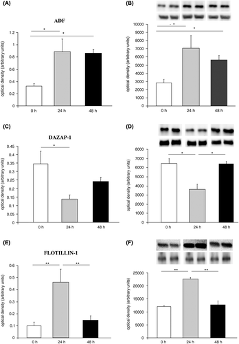
Table I. Protein levels (mean ± SD) of spots with significant differential expression at 0 (controls), 24, and 48 hours after serotonin treatment in Aplysia abdominal ganglia. Relative protein expression (arbitrary units) resulting from software-assisted quantification of 2-DE gels is given.
Table II. Results of statistical evaluation of spots differentially expressed in Aplysia abdominal ganglia 0 (controls), 24, and 48 hours after serotonin treatment. Relative protein expression resulting from software-assisted quantification of 2-DE gels was subjected to statistical analysis (one-way ANOVA followed by Bonferroni post hoc tests).
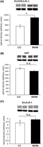
Figure 3. Protein levels of Flotillin-1 are increased after spatial learning in the mouse hippocampus. Levels of (A) Flotillin-1, (B) ADF, and (C) DAZAP-1 in hippocampal tissue of mice trained in the Morris water maze (MWM) and yoked controls (CO) (n = 6–8 per group). Inserts depict representative images of original Western blots of target proteins and β-tubulin loading control, respectively. All data display optical densities as mean ± SEM. *P < 0.05; n.s. = not significant.