Figures & data
Figure 1. The collagen synthesis pathway.
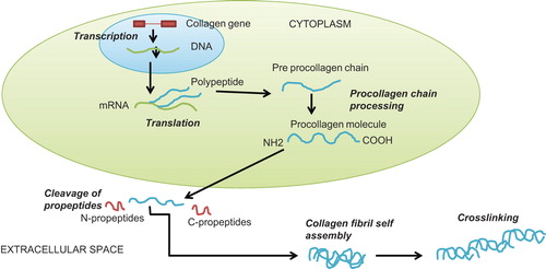
Table I. Detailed patient characteristics.
Table II. Characteristics of idiopathic myocardial fibrosis (IMF) cases and controls.
Figure 2. Immunohistochemical staining of PINP in myocardium of idiopathic fibrosis case (A) and in control case (B). Magnification × 200. The amount of PINP (stained brown) was increased in the fibrotic tissue of the IMF case in comparison with control sample.
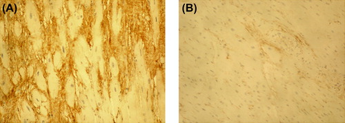
Figure 3. Immunohistochemical staining of ICTP in myocardium of idiopathic fibrosis case (A) and in control case (B). Magnification × 200. ICTP reflecting collagen type I degradation did not differ between the IMF case and control myocardium.
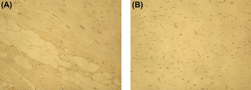
Figure 4. Immunohistochemical staining of PIIINP in myocardium of lamin A/C mutation case (A) and in control case (B). Magnification × 200. The amount of PIIINP (stained brown) was slightly increased in the IMF case in comparison with the control case.
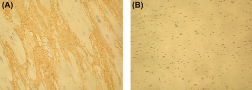
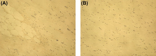
Figure 5. Immunohistochemical staining of IIINTP in myocardium of idiopathic fibrosis case (A) and in control case (B). Magnification × 200. The amount of IIINTP did not differ between the IMF case and control myocardium.