Figures & data
Figure 1. Schematic representation of the HLA locus. Scheme shows the different genes located in the class-I and class-II region of the HLA locus. Genes that encode classical HLA class I and class II molecules are indicated in black.
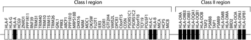
Figure 2. Schematic representation of the progression from healthy to ACPA+ RA. The HLA class II locus associates with the progression from ACPA positivity to ACPA+ RA. During this ‘second hit’ the ACPA response matures as shown by isotype expansion, increased ACPA levels, and epitope spreading. Together it supports a role for the HLA class II locus in the maturation of ACPA.
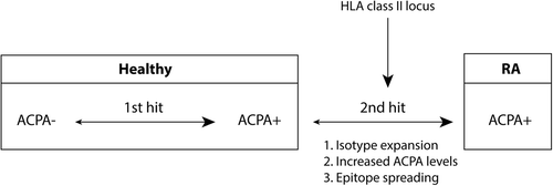
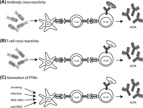
Figure 3. Models explaining loss of tolerance in RA. A: Infection and subsequent citrullination of microbial proteins results in the activation of microbe-directed T-cells. These microbe-directed T-cells can provide help to B-cells specific for a citrullinated microbial protein resulting in the production of ACPA cross-reactive to citrullinated self-proteins. Microbial proteins could become citrullinated by human or by bacterial PADI enzymes. B: Infection with microbes that have proteins with a high degree of similarities with self-protein could activate self-reactive T-cells. These self-reactive T-cells can subsequently provide help to B-cells specific for citrullinated self-proteins resulting in the production of ACPA. C: The amount of citrullinated proteins could be enhanced by smoking, infection with PADI-containing bacteria, polymorphisms within the PADI locus, or by anti-PADI antibodies. Enhanced citrullination of self-proteins occurs by several causes. Presentation of citrullinated neo-antigens by HLA-SE molecules activates citrulline-directed CD4 + T-cells that can help ACPA-producing B-cells. In each of the models antigens indicated in white are self-derived and antigens indicated in grey are pathogen-derived.