2,190
Views
50
CrossRef citations to date
0
Altmetric
Review Article

Inherited bone marrow failure syndromes in adolescents and young adults

, , &
Pages 353-363 | Received 13 Dec 2013, Accepted 11 Apr 2014, Published online: 03 Jun 2014

Figures & data

Table I. Examples of inherited bone marrow failure syndromes (IBMFS).

Figure 1. Phenotypic features of Fanconi anemia (FA). (A) Hypoplastic thumbs in an 11-year-old. (B) Hypopigmented (white arrow) and hyperpigmented (black arrows) skin lesions on the trunk of an adolescent. Photograph courtesy of Susan J. Bayliss, MD. (C) Progressive aplastic anemia in a 20-year-old with FA. Most of the marrow space is occupied by fat.

Figure 1. Phenotypic features of Fanconi anemia (FA). (A) Hypoplastic thumbs in an 11-year-old. (B) Hypopigmented (white arrow) and hyperpigmented (black arrows) skin lesions on the trunk of an adolescent. Photograph courtesy of Susan J. Bayliss, MD. (C) Progressive aplastic anemia in a 20-year-old with FA. Most of the marrow space is occupied by fat.

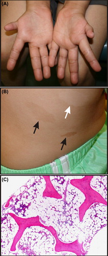
Table II. Congenital and developmental anomalies associated with Fanconi anemia (FA) (Citation3,Citation95).

Figure 2. Phenotypic features of dyskeratosis congenita (DC). (A) Nail dystrophy (arrow) in an adolescent. (B) Oral leukoplakia in an adult. (C) Skin changes in an adult. Hypopigmented lesions and the typical reticular rash of DC are evident on the upper chest (black arrow). Hypopigmentation is more pronounced in the sun-damaged skin of the neck (white arrow). (D) and (E) Pulmonary fibrosis on chest radiograph and CT scan of an adult. Photographs courtesy of Susan J. Bayliss, MD, and William H. McAlister, MD.

Figure 2. Phenotypic features of dyskeratosis congenita (DC). (A) Nail dystrophy (arrow) in an adolescent. (B) Oral leukoplakia in an adult. (C) Skin changes in an adult. Hypopigmented lesions and the typical reticular rash of DC are evident on the upper chest (black arrow). Hypopigmentation is more pronounced in the sun-damaged skin of the neck (white arrow). (D) and (E) Pulmonary fibrosis on chest radiograph and CT scan of an adult. Photographs courtesy of Susan J. Bayliss, MD, and William H. McAlister, MD.

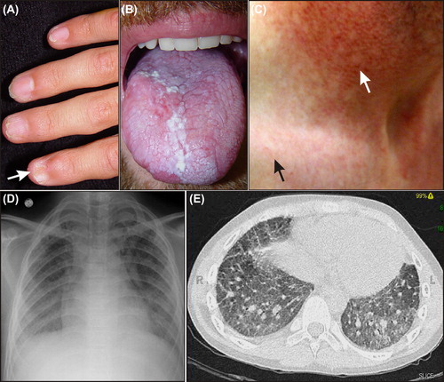
Table III. Prevalence of clinical features in patients with classic dyskeratosis congenita (DC) (Citation3,Citation22,Citation95).

Figure 3. Phenotypic features of Diamond–Blackfan anemia (DBA). (A) Abnormal thumb in an adult with an RPL11 mutation. (B) Muscular ventricular septal defect. Note the blood flow across the defect. LV = left ventricle; RV = right ventricle. Echocardiogram courtesy of Patrick Jay, MD, PhD.

Figure 3. Phenotypic features of Diamond–Blackfan anemia (DBA). (A) Abnormal thumb in an adult with an RPL11 mutation. (B) Muscular ventricular septal defect. Note the blood flow across the defect. LV = left ventricle; RV = right ventricle. Echocardiogram courtesy of Patrick Jay, MD, PhD.

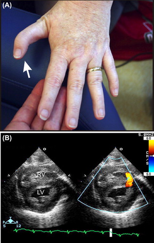
Table IV. Congenital and developmental anomalies associated with Diamond–Blackfan anemia (DBA) (Citation35,Citation96–98).

Figure 4. Phenotypic features of thrombocytopenia with absent radii (TAR) syndrome. (A) Absence of the radius. (B) Late-onset MDS in an adolescent with TAR syndrome (see Vignette 6). Radiograph courtesy of William H. McAlister, MD.

Figure 4. Phenotypic features of thrombocytopenia with absent radii (TAR) syndrome. (A) Absence of the radius. (B) Late-onset MDS in an adolescent with TAR syndrome (see Vignette 6). Radiograph courtesy of William H. McAlister, MD.

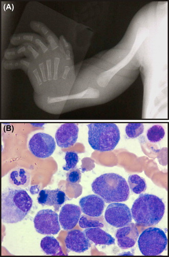
Table V. Congenital and developmental anomalies associated with Shwachman–Diamond syndrome (SDS) (Citation70,Citation99,Citation100).

Figure 5. Phenotypic features of Shwachman–Diamond syndrome (SDS). (A) MRI showing fatty infiltration of the pancreas (white arrow). (B) Metaphyseal dysostosis (black arrows). Images courtesy of William H. McAlister, MD.

Figure 5. Phenotypic features of Shwachman–Diamond syndrome (SDS). (A) MRI showing fatty infiltration of the pancreas (white arrow). (B) Metaphyseal dysostosis (black arrows). Images courtesy of William H. McAlister, MD.

Reprints and Corporate Permissions

Please note: Selecting permissions does not provide access to the full text of the article, please see our help page How do I view content?

To request a reprint or corporate permissions for this article, please click on the relevant link below:

Academic Permissions

Please note: Selecting permissions does not provide access to the full text of the article, please see our help page How do I view content?

Obtain permissions instantly via Rightslink by clicking on the button below:

If you are unable to obtain permissions via Rightslink, please complete and submit this Permissions form. For more information, please visit our Permissions help page.