Figures & data
Figure 1. Experimental paradigm and pupillary and amygdala responses to the aversive stimulus. A: Training and test phase: The training phase (left) consisted of several explicitly unpaired (bottom row) or paired (top row) presentations of the CS and the US and was followed by a period of rest (middle) during which the structural MRI images were acquired. The test phase (right) consisted of five presentations of the CS alone. B: Presentation of the screams during the training phase led to significant pupillary dilation in safety and fear conditioning groups (*P < 0.05) which was comparable among the two groups (P > 0.05). C: Activity in the left and right amygdala in both groups (average effect of condition from both groups) (Montreal Neurological Institute (MNI) co-ordinates: −18, −4, −14, k = 226, t score = 5.30, P < 0.05, FWE corrected; and 20, 0, −14, k = 244, t score = 5.15, P < 0.05, FWE corrected).
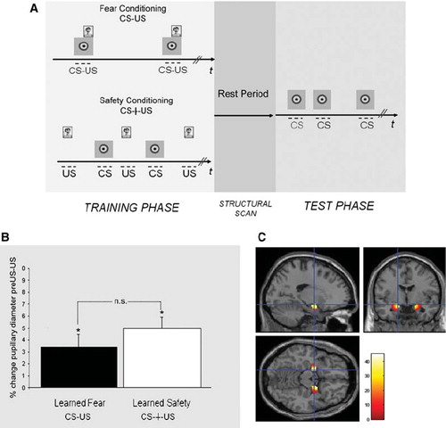
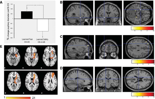
Figure 2. Pupillary and neural responses to the conditioned stimulus in the test phase. A: Percentage change in pupillary diameter during the presentation of the first CS (*P < 0.05) demonstrates pupillary constriction in the learned safety group and pupillary dilation in the learned fear group. B: A cluster of differential activation in the left amygdala between safety and fear trained subjects in response to the CS is shown on a standard brain (Montreal Neurological Institute (MNI) co-ordinates: −28, 0, −16, k = 8, t score = 3.56, P < 0.05, FWE corrected). For display purposes, activity clusters are shown at P < 0.005, uncorrected in B and C. C: Differential activation in the left dlPFC between safety and fear trained groups in response to the CS (MNI: −30, 52, 32, k = 9, t score = 3.97, P < 0.001 uncorrected). D. Differential activation in the left caudate between safety and fear trained groups in response to the CS (MNI: −16, −18, 20, k = 12, t score = 2.11, P = 0.021 uncorrected). E. Group-based probabilistic tractography map between the amygdala and the dlPFC.
Supplemental Table I. Regions with greater activity (at P < 0.001, uncorrected) in the safety than in the fear conditioned group (whole brain analysis).
Supplemental Table II. Number of streamlines (waytotals) for each subject.