Figures & data
Table I. Demographic characteristics and laboratory findings in type 1 and type 2 diabetic patients divided into groups with normal and enlarged left atrium volume indexed to body surface area (LAVI).
Figure 1. Prevalence of increased left atrium volume index (LAVI) in different subgroups of patients: (Citation1) normotensive patients (NT) with normal peak early transmitral jet velocity to mitral annulus velocity ratio (E/E’), (Citation2) hypertensive patients (HT) with normal E/E’ and (Citation3) patients with increased E/E’ (presence of left ventricular diastolic dysfunction) according to type of diabetes.
Table II. Echocardiographic findings in type 1 and type 2 diabetic patients divided into groups with normal and increased left atrium volume index (LAVI).
Table III. Correlates of larger left atrium volume index (LAVI) in the total population and in groups of patients with type 1 and type 2 diabetes.
Figure 1. Relationship between left atrium volume index (LAVI) and peak early transmitral jet velocity to mitral annulus velocity ratio (E/E’) in type 1 and type 2 diabetic patients.
Table IV. Independent predictors of left atrium volume index (LAVI) in type 1 and type 2 diabetic patient groups identified in multivariate linear regression analyses.
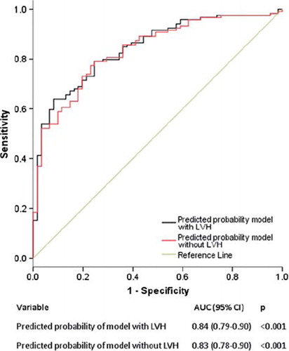
Figure 1. Enlarged left atrium volume index (LAVI) was associated with left ventricular (LV) diastolic dysfunction independent of presence of LV hypertrophy. Receiver operating characteristic (ROC) curve showing predicted probability of the multivariate logistic regression model with and without LV hypertrophy in the model.