634
Views
18
CrossRef citations to date
0
Altmetric
Laboratory Study

apoE Expression in Glomerulus and Correlation with Glomerulosclerosis Induced by Adriamycin in Rats

, , , , &
Pages 348-354 | Received 06 Nov 2010, Accepted 27 Jan 2011, Published online: 14 Mar 2011

Figures & data

Table 1. Parameters in two groups (mean ± SD)

Figure 1. Glomerular morphology was normal in sham group (A1: 9 weeks; A2: 13 weeks; HE). Proliferation occurring in the majority of glomerular mesangial cells and extracellular matrix in GS group, and degeneration of glomerular epithelial cells and infiltration of widespread mononuclear cells were shown (A3: 9 weeks; A4: 13 weeks; HE), especially in 13 weeks of GS group (A4). Representative samples of immunohistochemical staining for glomerular Col-IV (SHO: B1: 9 weeks, B2: 13 weeks; GS: B3: 9 weeks and B4: 13 weeks), FN (SHO: C1: 9 weeks, C2: 13 weeks; GS: C3: 9 weeks and C4: 13 weeks), α-SMA (SHO: D1: 9 weeks, D2: 13 weeks; GS: D3: 9 weeks and D4: 13 weeks), TGF-β1 (SHO: E1: 9 weeks, E2: 13 weeks; GS: E3: 9 weeks and E4: 13 weeks), and apoE (SHO: F1: 9 weeks, F2: 13 weeks; GS: F3: 9 weeks and F4: 13 weeks) were observed in all groups. Sham group (B1, B2, C1, C2, D1, D2, E1, E2, F1, and F2): positive staining (in brown) was faint in glomerulus, glomerular endothelial cells, glomerular basement membrane, mesangial cells, and visceral epithelial cells. GS group (B3, B4, C3, C4, D3, D4, E3, E4, F3, and F4): positive staining was strong in most glomerulus, glomerular endothelial cells, glomerular basement membrane, mesangial cells, and visceral epithelial cells, especially in those of 13 weeks (B4, C4, D4, E4, and F4). Magnification 400×.

Notes: SHO, sham operation group; GS, glomerulosclerosis model group; HE, hematoxylin and eosin; FN, fibronectin; α-SMA, α-smooth muscle actin; TGF-β1, transforming growth factor-β1; apoE, apolipoprotein E.

Figure 1. Glomerular morphology was normal in sham group (A1: 9 weeks; A2: 13 weeks; HE). Proliferation occurring in the majority of glomerular mesangial cells and extracellular matrix in GS group, and degeneration of glomerular epithelial cells and infiltration of widespread mononuclear cells were shown (A3: 9 weeks; A4: 13 weeks; HE), especially in 13 weeks of GS group (A4). Representative samples of immunohistochemical staining for glomerular Col-IV (SHO: B1: 9 weeks, B2: 13 weeks; GS: B3: 9 weeks and B4: 13 weeks), FN (SHO: C1: 9 weeks, C2: 13 weeks; GS: C3: 9 weeks and C4: 13 weeks), α-SMA (SHO: D1: 9 weeks, D2: 13 weeks; GS: D3: 9 weeks and D4: 13 weeks), TGF-β1 (SHO: E1: 9 weeks, E2: 13 weeks; GS: E3: 9 weeks and E4: 13 weeks), and apoE (SHO: F1: 9 weeks, F2: 13 weeks; GS: F3: 9 weeks and F4: 13 weeks) were observed in all groups. Sham group (B1, B2, C1, C2, D1, D2, E1, E2, F1, and F2): positive staining (in brown) was faint in glomerulus, glomerular endothelial cells, glomerular basement membrane, mesangial cells, and visceral epithelial cells. GS group (B3, B4, C3, C4, D3, D4, E3, E4, F3, and F4): positive staining was strong in most glomerulus, glomerular endothelial cells, glomerular basement membrane, mesangial cells, and visceral epithelial cells, especially in those of 13 weeks (B4, C4, D4, E4, and F4). Magnification 400×.Notes: SHO, sham operation group; GS, glomerulosclerosis model group; HE, hematoxylin and eosin; FN, fibronectin; α-SMA, α-smooth muscle actin; TGF-β1, transforming growth factor-β1; apoE, apolipoprotein E.

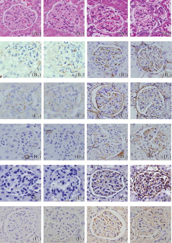
Table 2. Protein expression of apoE in glomerulus and mRNA expression of apoE in renal tissue of the two groups (mean ± SD)

Reprints and Corporate Permissions

Please note: Selecting permissions does not provide access to the full text of the article, please see our help page How do I view content?

To request a reprint or corporate permissions for this article, please click on the relevant link below:

Academic Permissions

Please note: Selecting permissions does not provide access to the full text of the article, please see our help page How do I view content?

Obtain permissions instantly via Rightslink by clicking on the button below:

If you are unable to obtain permissions via Rightslink, please complete and submit this Permissions form. For more information, please visit our Permissions help page.