Figures & data
Figure 1. Malondialdehyde (MDA) level in the renal tissue of sham surgery (SS), renal ischemia (RI), renal ischemia-reperfusion (RIR), and mirtazapine + renal ischemia-reperfusion (MRIR) groups. The results are expressed as mean ± SEM.
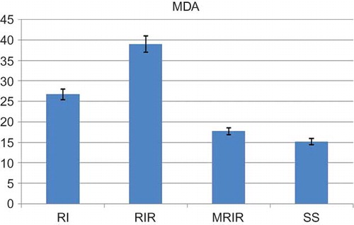
Figure 2. Myeloperoxidase (MPO) activities in kidneys of sham surgery (SS), renal ischemia (RI), renal ischemia-reperfusion (RIR), and mirtazapine + renal ischemia-reperfusion (MRIR) groups. The results are expressed as mean ± SEM.
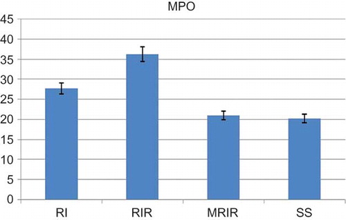
Figure 3. Glutathione (GSH) level in the renal tissue of sham surgery (SS), renal ischemia (RI), renal ischemia-reperfusion (RIR), and mirtazapine + renal ischemia-reperfusion (MRIR) groups. The results are expressed as mean ± SEM.
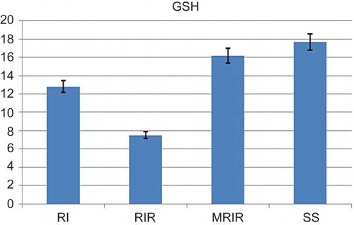
Figure 4. Glutathione S-transferase (GST) activities in kidneys of sham surgery (SS), renal ischemia (RI), renal ischemia-reperfusion (RIR), and mirtazapine + renal ischemia-reperfusion (MRIR) groups. The results are mean ± SEM.
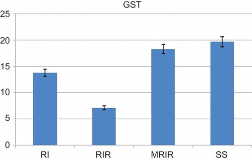
Table 1. The MDA and GSH levels and MPO and GST activities detected in contralateral kidneys of RICL, RIRCL, and MRIRCL groups.
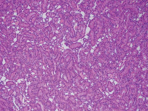
Figure 5. The histopathological examination of the renal tissue of sham surgery (SS) group.
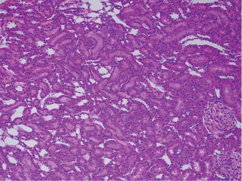
Figure 6. The histopathological examination of the renal tissue of renal ischemia (RI) group. Hemorrhage (arrow), but no tubular injury, was observed in interstitial area.
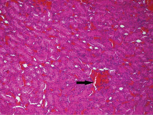
Figure 7. The histopathological examination of the renal tissue of renal ischemia-reperfusion (RIR) group. Remarkable tubular epithelial swelling (A, Arrow-1), tubular epithelial necrosis (A, Arrow-2), hyaline cast accumulation in dilated tubules (A, Arrow-3), and loss of microvillus (B, Arrow-1) were observed.
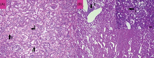
Figure 8. The histopathological examination of the renal tissue of mirtazapine + renal ischemia-reperfusion (MRIR) group. Mild swelling in tubular epithelial cells (A, Arrow) and mild hyaline cast accumulation (B, Arrow-1) were observed. Microvilli structures in proximal tubules were observed to be preserved (B, Arrow-2).
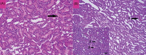
Figure 9. The histopathological examination of the renal tissue of mirtazapine + renal ischemia-reperfusion contralateral kidney (MRIRCL) group.
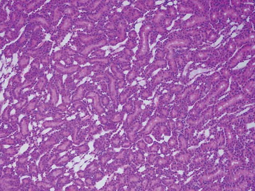
Figure 10. The histopathological examination of the renal tissue of renal ischemia-reperfusion contralateral kidney (RIRCL) group. Mild hemorrhage in interstitial area (Arrow-1) and tubular hyaline cast accumulation (Arrow-2) were observed. Microvilli structures in proximal tubular epithelial cells appeared to be preserved (Arrow-3).
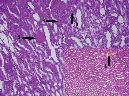
Figure 11. The histopathological examination of the renal tissue of renal ischemia contralateral kidney (RICL) group.