Figures & data
Table 1. Primer sequences of amplified genes.
Table 2. Differences of body weight (BW), 24 h urine protein, and urine NAG between different groups ().
Table 3. Differences of Scr, BUN, Hb, and RBC between different groups ().
Figure 1. Hematoxylin–eosin staining in rat renal tissue samples at week 21 (×400). (A) There was no significant histological abnormality in the control group. (B) Degenerated, necrotic, and sloughed tubule epithelial cells and an exposed basement membrane were seen, and some of the tubule structures were atrophied and lost. (C) In Cozaar group, only local mild tubular epithelial lesions were observed.
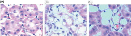
Figure 2. Masion staining in rat renal tissue samples at week 21 (×400). (A) There was no fibrosis of the interstitium in the control group. (B) A moderate fibrosis of the interstitium was seen in the model group. (C) A less severe interstitial fibrosis was observed in Cozaar group.
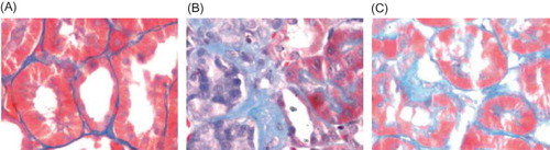
Figure 3. Electron microscopy of glomerulus at week 21 (×4200). (A) The ultrastructure of intact glomerulus was seen in the control group. (B) An ischemic shrinkage of the glomerular basement membrane was found in the model group. (C) A less severe ischemic shrinkage of the glomerular basement membrane was observed in Cozaar group.
Figure 4. Electron microscopy of rat renal tubule at week 21 (×11,500). (A) The normal tubule epithelial cells and basement membrane were seen in the control group. (B) Thickening and collapse of the tubule basement membrane were found in the model group. (C) A mild thickening and collapse of the tubule basement membrane was observed in Cozaar group.
Figure 5. Electron microscopy of rat renal interstitium at week 21 (×11,500). (A) The normal ultrastructure of renal interstitium was seen in the control group. (B) A significant amount of fasciculation of collagen fibers at the affected interstitium was found in the model group. (C) Less fasciculations of collagen fibers at the affected interstitium were observed in Cozaar group.
Figure 6. BMP-7 expression in rat renal tissue at week 21 (×250). (A) BMP-7 was largely expressed in the renal tubular epithelial cells and renal interstitium, especially in the medulla area in the control group. (B) A decreased BMP-7 expression was found in the renal tubular epithelial cells and renal interstitium. (C) BMP-7 expression in the renal tissue was significantly increased in Cozaar group.
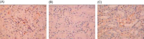
Table 4. Differences of relative positive area of BMP-7, caspase-3, and CD34 expressions in rat renal tissue between different groups ().
Figure 7. Caspase-3 expression in rat renal tissue (×250). (A) Caspase-3 was less expressed in the renal tubular interstitium in the control group. (B) An increased caspase-3 expression was found in the renal interstitium in the model group. (C) Caspase-3 expression in the renal tissue was significantly decreased in Cozaar group.
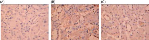
Figure 8. CD34 expression in rat renal tissue (×250). (A) CD34 was largely expressed in the renal tubular interstitium in the control group. (B) A decreased CD34 expression was found in the renal interstitium in the model group. (C) CD34 expression in the renal tissue was significantly increased in Cozaar group as compared with the model group.
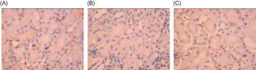
Table 5. Differences of Ang-1, Ang-2, Tie-2, and VEGF mRNA expressions between different groups ().