Figures & data
Table 1. Primer sequence.
Table 2. Positive rates of the apoptotic proteins (mean ± SD; n = 10).
Figure 1. Histologic changes in the kidney at the end of 48 h of reperfusion. Tissues were stained with hematoxylin and eosin. Shown are representative histological specimens from the control group (A); renal I/R + NS group (B); and I/R + NGAL group (C). Magnification ×100.
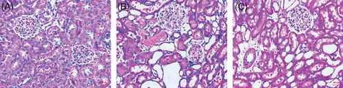
Figure 2. Representative photomicrographs of the outer medulla illustrate apoptotic nuclei. The control group rarely showed apoptotic nuclei (A, magnification ×100); rats subjected to I/R + NS showed TUNEL-positive cells (B, magnification ×100; D, magnification ×400); rats subjected to I/R + NGAL showed a smaller degree of TUNEL-positive cells (C, magnification ×100; E, magnification ×400). Arrows indicate TUNEL-positive cells. *p, **p < 0.05, versus I/R + NS group.
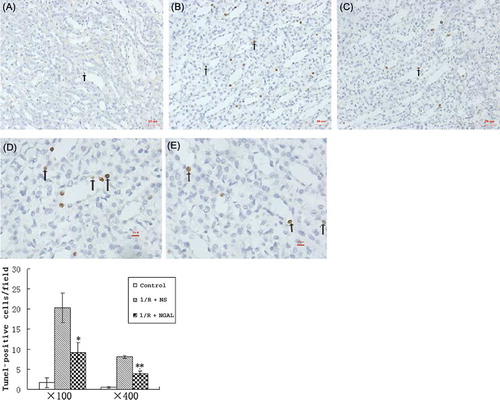
Figure 3. The mRNA levels of (A) Fas and (B) Bcl-2 in the kidney at the end of 48 h of reperfusion. *p < 0.05, versus I/R + NS group.
Figure 4. Immunohistochemical staining of Fas in control group (A), I/R + NS group (B), and I/R + NGAL (C); Fas staining was strong on tubular epithelial cell membrane. Bcl-2 in control group (D), I/R + NS group (E), and I/R + NGAL (F); Bcl-2 staining was strong on tubular epithelial cell plasma. Magnification ×100. The magnifications b, c, e, and f were taken from the insert frames.
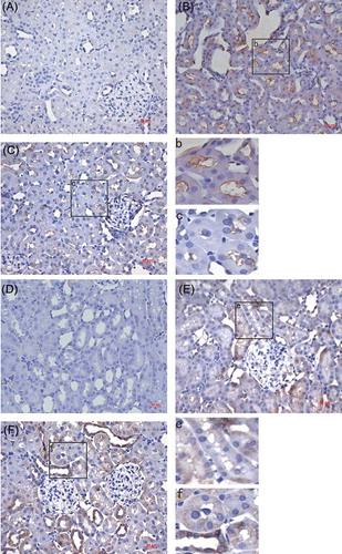
Figure 5. Immunoblot analysis of Fas and Bcl-2 proteins (A). The density of band was quantified. Values presented are ratios of Fas or Bcl-2 to beta-actin, which was used as an equal protein loading marker (B and C). Results are presented as mean ± SD (n = 5); *p < 0.05, versus IR + NS group; **p < 0.05, versus IR + NS group.