Figures & data
Figure 1. Chest CT scan.
Note: A solitary nodule (32 × 30 mm) in the lingual lobe (arrow).
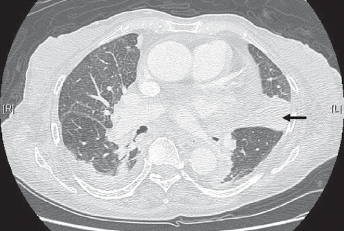
Figure 2. Renal biopsy.
Note: Congo-red stain reveals apple-green birefringence under polarized light.
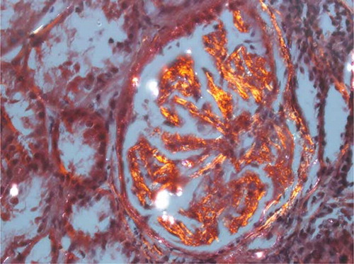
Figure 3. Lung biopsy.
Note: Congo-red stain with apple-green birefringence under polarized light.
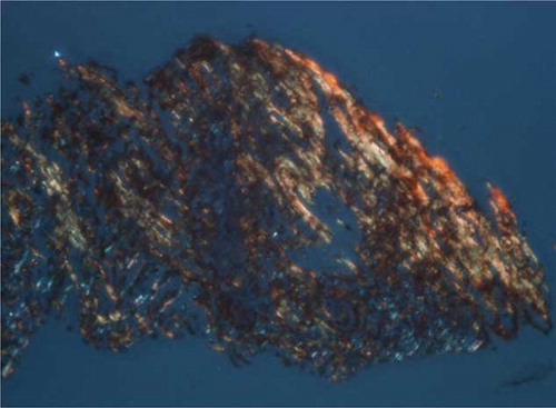
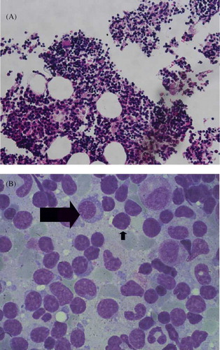
Figure 4. Bone marrow biopsy (A) predominantly mature B-lymphocytes in myeloid (hematoxylin and eosin stain, magnification ×400). (B) Increased aggregation of lymphocytes (short arrow) and plasma cells (long arrow).