Figures & data
Table 1. Biopsy findings and diagnosis of groups 1 and 2.
Figure 1. Photomicrograph showing exfoliated esophageal squamous epithelial cells with herpes simplex nuclear inclusions (arrow), hematoxylin eosin stain, 400×.
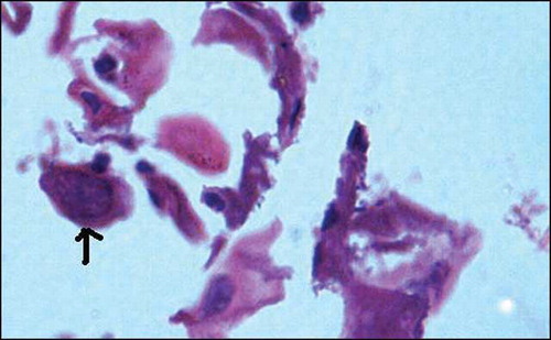
Figure 2. Duodenal mucosal biopsy showing strongyloides larvae within glands (arrow), hematoxylin eosin stain, 200×.
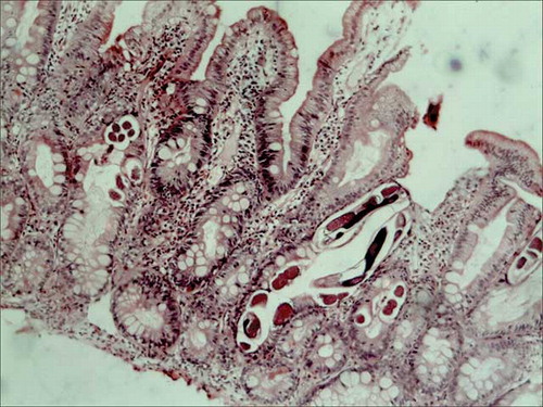
Figure 3. Photomicrograph showing infarcted colonic mucosa in ischemic colitis with adjacent mucosal hemorrhage, hematoxylin eosin stain, 200×.
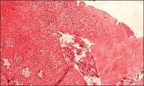
Figure 4. Colonic mucosal biopsy showing CMV inclusions in mucosal capillary endothelium (arrow), hematoxylin eosin stain, 400×.
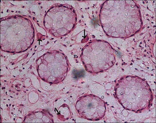
Figure 5. Colonic mucosal biopsy showing marked crypt architectural distortion, branching and clustering of crypts, intervening widely spaced lamina propria shows edema and inflammation, features reminiscent of idiopathic inflammatory bowel disease, hematoxylin eosin stain, 200×.
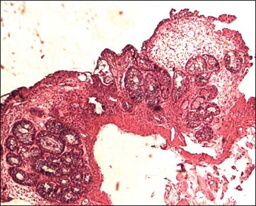
Figure 6. Colonic mucosal biopsy showing significant reduction in crypts, consistent with MMF-related toxic changes, hematoxylin eosin stain, 200×.
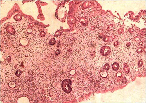
Table 2. MMF-related colitis.